
Cardiovascular System
Identification and Nursing Management of Congenital Malformations
Comprehensive nursing notes for education and clinical reference
Contents
Overview of Congenital Heart Defects
Congenital heart defects (CHDs) are structural problems arising from abnormal formation of the heart or major blood vessels that are present at birth. They are the most common type of birth defect, affecting approximately 1% of live births worldwide.
Key Facts
- CHDs affect nearly 1 in every 100 babies born each year in the United States
- About 25% of babies with a CHD have a critical CHD that requires surgery or other procedures within the first year of life
- Congenital heart defects are responsible for about 30% of deaths due to birth defects
- Survival rates have significantly improved with advances in diagnosis and treatment
Congenital heart defects result from disruptions in the normal process of heart development during pregnancy. They can range from simple conditions with no symptoms to complex problems causing severe, life-threatening symptoms. Modern treatments have significantly improved outcomes, allowing most children with CHDs to survive to adulthood and lead productive lives.
Nursing Perspective
As nurses caring for patients with CHDs, understanding the anatomical and physiological implications of each defect is crucial for effective assessment, care planning, and patient/family education. The primary nursing goals focus on symptom management, promoting growth and development, ensuring proper nutrition, preventing complications, and providing comprehensive support to families.
Embryology and Development
Understanding the embryological development of the heart is essential for comprehending how congenital heart defects occur. During normal heart development, the heart forms from a simple tube that loops and divides to create the four chambers and major blood vessels.
Timeline of Heart Development
- Day 15-16: Cardiogenic area appears
- Day 18-19: Heart tube forms
- Day 21-23: Heart begins to beat
- Day 22-23: Heart tube begins to loop
- Week 4-5: Formation of chambers begins
- Week 5-6: Development of atrial and ventricular septa
- Week 5-8: Formation of the great vessels
- Week 8: Basic heart structure complete
Critical Periods for CHD Development
Congenital heart defects typically develop during critical periods when specific heart structures are forming. Disruptions during these periods can lead to specific defects:
- Weeks 3-4: Heart tube looping defects
- Weeks 4-6: Septal defects and valve defects
- Weeks 5-7: Outflow tract defects
- Weeks 6-8: Great vessel defects

Figure 1: Embryonic Heart Development and Circulation Patterns
Fetal Circulation vs. Postnatal Circulation
Fetal circulation differs from postnatal circulation in several important ways:
- In fetal circulation, oxygenated blood from the placenta bypasses the lungs through the foramen ovale and ductus arteriosus
- At birth, the lungs expand, pulmonary vascular resistance decreases, and systemic vascular resistance increases
- These changes lead to functional closure of the foramen ovale and ductus arteriosus
- Failure of these transitions can lead to persistent fetal circulation patterns and associated defects
Types of Congenital Heart Defects
Congenital heart defects are typically classified into two major categories based on their hemodynamic effects and clinical presentation: acyanotic and cyanotic defects.
Classification Mnemonic: “CASH Flow”
Cyanotic vs. Acyanotic
Shunting direction (Left-to-Right or Right-to-Left)
Hemodynamic effects
Flow patterns and oxygenation changes
| Parameter | Acyanotic Defects | Cyanotic Defects |
|---|---|---|
| Shunt Direction | Usually Left-to-Right | Right-to-Left |
| Oxygenation | Normal arterial oxygen saturation | Decreased arterial oxygen saturation |
| Clinical Presentation | Heart failure, poor growth, respiratory infections | Central cyanosis, hypoxic spells, clubbing |
| Common Examples | ASD, VSD, PDA, Coarctation of aorta | TOF, TGA, Tricuspid Atresia, TAPVC |
| Pathophysiology | Increased pulmonary blood flow | Decreased pulmonary blood flow or mixing of oxygenated and deoxygenated blood |
Acyanotic Defects
Acyanotic defects are characterized by a left-to-right shunt, where oxygenated blood is redirected from the systemic circulation back to the pulmonary circulation. These defects usually cause volume overload but do not typically cause cyanosis initially.
Acyanotic Defects Mnemonic: “PAVS”
Patent Ductus Arteriosus
Atrial Septal Defect
Ventricular Septal Defect
Stenotic lesions (Aortic/Pulmonary Stenosis, Coarctation of Aorta)
1. Atrial Septal Defect (ASD)
Definition: A hole in the wall (septum) between the heart’s upper chambers (atria).
Physiology: Left-to-right shunting of blood across the atrial septum, leading to right atrial and ventricular volume overload and increased pulmonary blood flow.
Types:
- Secundum ASD (most common, located in center of septum)
- Primum ASD (located in lower portion of septum)
- Sinus venosus ASD (located near junction with SVC or IVC)
Clinical Features:
- Often asymptomatic in children
- Soft, systolic ejection murmur at upper left sternal border
- Fixed splitting of second heart sound (S2)
- May develop right ventricular failure in adulthood

Figure 2: Atrial Septal Defect (ASD) showing communication between left and right atria
Nursing Tip
For ASD patients, auscultate for fixed splitting of S2 that doesn’t change with respiration. This is a key diagnostic clue that distinguishes ASD from normal physiologic splitting.
2. Ventricular Septal Defect (VSD)
Definition: A hole in the wall (septum) between the heart’s lower chambers (ventricles).
Physiology: Left-to-right shunting across the ventricular septum, causing increased pulmonary blood flow and risk of pulmonary hypertension if left untreated.
Types:
- Perimembranous VSD (most common, 80%)
- Muscular VSD (located in the muscular portion)
- Inlet VSD (near AV valves)
- Outlet/Supracristal VSD (beneath the semilunar valves)
Clinical Features:
- Harsh holosystolic murmur at left lower sternal border
- Symptoms vary with defect size
- Small VSDs may be asymptomatic
- Large VSDs cause heart failure, poor weight gain, and recurrent respiratory infections

Figure 3: Ventricular Septal Defect (VSD) showing communication between ventricles
Clinical Alert
Large VSDs require close monitoring for signs of pulmonary hypertension. Watch for increased respiratory effort, right ventricular heave, and decreased exercise tolerance, which may indicate development of pulmonary vascular disease (Eisenmenger syndrome).
3. Patent Ductus Arteriosus (PDA)
Definition: Persistent patency of the ductus arteriosus, which normally closes shortly after birth.
Physiology: Continued blood flow from the aorta to the pulmonary artery, increasing pulmonary blood flow and left heart volume load.
Risk Factors:
- Prematurity (most common)
- Maternal rubella infection during pregnancy
- High altitude birth
- Genetic factors
Clinical Features:
- Continuous machine-like murmur (“machinery murmur”) at left upper sternal border/infraclavicular area
- Bounding pulses with wide pulse pressure
- In large PDAs: heart failure, poor growth, respiratory symptoms

Figure 4: Patent Ductus Arteriosus (PDA) showing persistent connection between aorta and pulmonary artery
Nursing Tip
When assessing premature infants with PDA, compare pre- and post-ductal oxygen saturations. A significant difference (>3-5%) may indicate a hemodynamically significant PDA that requires intervention.
4. Coarctation of the Aorta (CoA)
Definition: Narrowing of the aorta, usually occurring just distal to the left subclavian artery (juxtaductal).
Physiology: Causes obstruction to left ventricular outflow, leading to increased afterload, left ventricular hypertrophy, and decreased blood flow to the lower body.
Types:
- Preductal (proximal to ductus arteriosus)
- Juxtaductal (at the level of ductus arteriosus) – most common
- Postductal (distal to ductus arteriosus)
Clinical Features:
- Hypertension in upper extremities with normal/low blood pressure in lower extremities
- Diminished or delayed femoral pulses compared to radial pulses
- Systolic ejection murmur best heard at left interscapular area
- May present with heart failure in infants

Figure 5: Coarctation of the Aorta showing narrowing of the aortic arch
Assessment Pearl
Always compare blood pressure measurements in all four extremities for suspected CoA. A systolic pressure difference of >20 mmHg between upper and lower extremities is significant for coarctation.
Cyanotic Defects
Cyanotic defects are characterized by right-to-left shunting of blood, where deoxygenated blood bypasses the lungs and enters the systemic circulation, resulting in central cyanosis and hypoxemia.
Cyanotic Defects Mnemonic: “The 5 T’s”
Tetralogy of Fallot
Transposition of the Great Arteries
Tricuspid Atresia
Total Anomalous Pulmonary Venous Connection
Truncus Arteriosus
1. Tetralogy of Fallot (TOF)
Definition: A combination of four defects: ventricular septal defect (VSD), pulmonary stenosis, right ventricular hypertrophy, and overriding aorta.
Physiology: Right-to-left shunting through the VSD due to pulmonary stenosis, causing reduced pulmonary blood flow and systemic cyanosis.
Clinical Features:
- Progressive cyanosis that worsens with crying or exertion
- Hypercyanotic (“tet”) spells – acute episodes of increased cyanosis and dyspnea
- Harsh systolic ejection murmur at left upper sternal border
- Squatting position to relieve symptoms (increases systemic vascular resistance)
- Clubbing of fingers and toes in older children
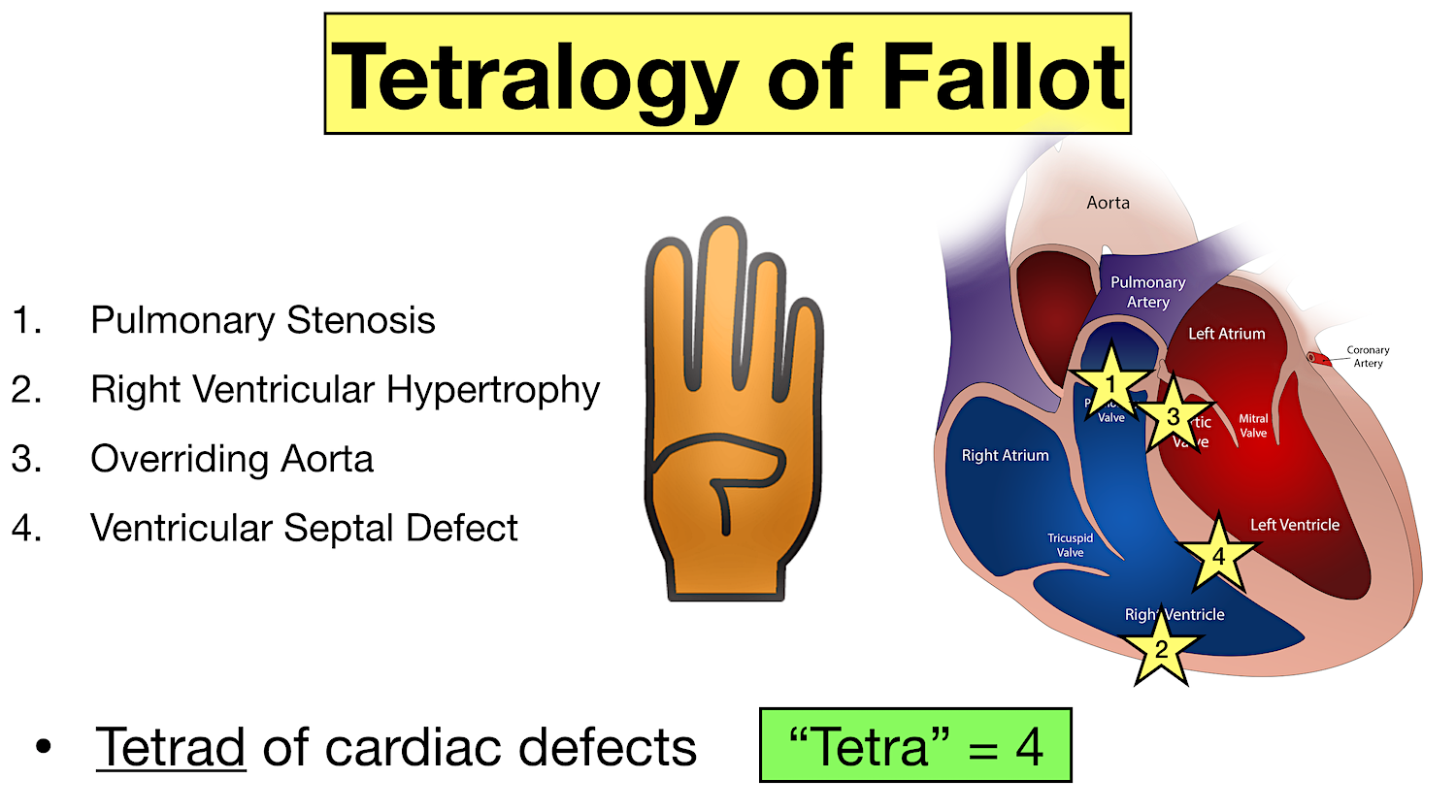
Figure 6: Tetralogy of Fallot showing the four characteristic defects
Emergency Management
For hypercyanotic (“tet”) spells, follow the “KNEE” approach:
- Knee-chest position or holding infant with knees to chest
- No crying (keep calm, provide comfort)
- Ensure oxygenation (administer oxygen)
- Emergency medications (morphine or phenylephrine if needed)
2. Transposition of the Great Arteries (TGA)
Definition: The aorta arises from the right ventricle and the pulmonary artery from the left ventricle, creating two separate parallel circulations.
Physiology: Deoxygenated blood returns to the body without being oxygenated in the lungs, while oxygenated blood returns to the lungs without delivering oxygen to the body. Survival depends on mixing via other defects (ASD, VSD, PDA).
Clinical Features:
- Severe cyanosis present from birth
- Minimal or no heart murmur if no associated defects
- Tachypnea and respiratory distress
- Hypoxemia not responsive to oxygen therapy
- Rapid deterioration without intervention
Figure 7: Transposition of the Great Arteries showing reversed connections of the great vessels
Nursing Tip
TGA is a surgical emergency. Prior to surgery, maintaining ductal patency with prostaglandin E1 (PGE1) infusion is critical for survival. Closely monitor for apnea (a side effect of PGE1) and have resuscitation equipment readily available.
3. Tricuspid Atresia
Definition: Complete absence or agenesis of the tricuspid valve, preventing direct blood flow from the right atrium to the right ventricle.
Physiology: Blood must flow from the right atrium to the left atrium through an ASD or patent foramen ovale. The right ventricle is typically hypoplastic, and a VSD is often present to allow some blood flow to the lungs.
Clinical Features:
- Cyanosis present from birth
- Systolic murmur due to associated VSD
- Single S2 heart sound
- Heart failure if increased pulmonary blood flow
- Hypoxic spells if decreased pulmonary blood flow
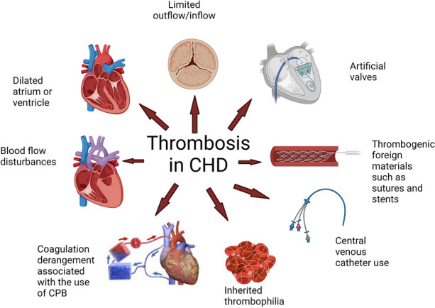
Figure 8: Tricuspid Atresia with absent tricuspid valve and hypoplastic right ventricle
Pathophysiology Key Point
Tricuspid atresia patients require a staged surgical approach (Fontan procedure), typically completing three surgeries by 2-4 years of age. These patients will have a single-ventricle physiology, with the left ventricle pumping blood to both the body and lungs.
Clinical Manifestations
The clinical presentation of congenital heart defects varies widely depending on the type and severity of the defect. Understanding the common manifestations is crucial for early recognition and timely intervention.
| System | Clinical Manifestations | Associated Defects |
|---|---|---|
| Cardiovascular |
|
Most CHDs, especially VSD, ASD, PDA, CoA |
| Respiratory |
|
Large left-to-right shunts (VSD, PDA), Complex cyanotic defects |
| Integumentary |
|
Cyanotic defects (TOF, TGA, Tricuspid Atresia) |
| Gastrointestinal |
|
CHDs with heart failure, Complex defects |
| Neurological |
|
Severe cyanotic defects, Defects with heart failure |
Age-Related Presentations
Neonatal Period (0-1 month)
- Cyanosis (TGA, TOF, Tricuspid Atresia)
- Respiratory distress
- Poor feeding
- Shock (HLHS, Critical CoA)
- Heart murmurs may not be present initially
Infancy (1-12 months)
- Heart failure symptoms (large VSD, PDA)
- Failure to thrive
- Recurrent respiratory infections
- Progressive cyanosis (TOF)
- Characteristic murmurs become apparent
Childhood (1-12 years)
- Exercise intolerance
- Growth delay
- Clubbing in cyanotic defects
- Squatting behavior (TOF)
- Some ASDs and small VSDs may be first detected
Red Flag Signs Requiring Immediate Attention
Neonates and Infants
- Central cyanosis not improving with oxygen
- Poor perfusion, weak pulses, or shock
- Respiratory rate >60/min with retractions
- Hepatomegaly with respiratory distress
- Sudden deterioration in the first week of life (ductal-dependent lesions)
Children
- Hypercyanotic (tet) spells
- Syncope or near-syncope with exertion
- Unexplained stroke or brain abscess
- Significant exercise limitation
- Chest pain with exertion
Nursing Observation Pearl
The timing of symptom onset can provide clues to the underlying defect:
- Immediate after birth: TGA, TAPVC, Pulmonary atresia
- Hours to days: Ductal-dependent lesions (HLHS, Critical CoA, Interrupted aortic arch)
- Days to weeks: Large VSD, PDA
- Months to years: ASD, small VSD, mild PS
Diagnosis and Assessment
Accurate diagnosis of congenital heart defects involves a combination of clinical assessment, diagnostic imaging, and specialized cardiac testing. Early diagnosis is crucial for timely intervention and improved outcomes.
History and Physical Examination
Key components of the initial assessment include:
- Family history: CHDs, genetic syndromes, maternal conditions
- Pregnancy history: Maternal infections, medication use, diabetes
- Birth history: Gestational age, birth weight, Apgar scores
- Growth parameters: Height, weight, head circumference
- Vital signs: Heart rate, respiratory rate, blood pressure (all 4 extremities)
- Cardiac examination: Heart sounds, murmurs, rhythm, pulses
- Respiratory examination: Work of breathing, retractions, crackles
- General appearance: Color, activity level, dysmorphic features
Laboratory Studies
- Pulse oximetry screening: Measures blood oxygen levels (routine in newborns)
- Arterial blood gases: Assess oxygenation and acid-base status
- Complete blood count: Evaluate for polycythemia in cyanotic defects
- Electrolytes: Monitor for imbalances with heart failure
- BNP/NT-proBNP: Biomarkers for heart failure
- Genetic testing: For associated syndromes (e.g., Down syndrome, DiGeorge syndrome)
Pulse Oximetry Screening
Critical congenital heart defect screening is performed on all newborns using pulse oximetry before discharge. A positive screen is:
- SpO2 <90% in right hand or either foot
- SpO2 of 90-94% in both right hand and either foot on repeated measurements
- Difference of >3% between right hand and foot
Diagnostic Imaging
Echocardiography
The gold standard non-invasive test for diagnosis of CHDs.
- Transthoracic echocardiography (TTE): Provides detailed images of cardiac structures, flow patterns, and function
- Fetal echocardiography: Allows prenatal diagnosis as early as 18-22 weeks gestation
- Transesophageal echocardiography (TEE): Used during interventions or when TTE views are limited
Chest X-ray
- Evaluates heart size and contour
- Assesses pulmonary vascularity (increased, decreased, normal)
- Identifies specific cardiac chamber enlargement
- Reveals associated lung pathology
Advanced Imaging
- Cardiac MRI: Provides detailed anatomical and functional assessment, especially for complex defects or extracardiac structures
- Cardiac CT: Offers rapid acquisition of images, useful for evaluating great vessels and coronary arteries
- 3D imaging: Enhanced visualization for complex defects and surgical planning
Cardiac Catheterization
- Provides hemodynamic data (pressures, oxygen saturations)
- Allows direct measurement of shunts and resistances
- Enables therapeutic interventions (balloon procedures, device closures)
- Evaluates coronary anatomy when needed
Other Diagnostic Tests
Electrocardiogram (ECG)
- Chamber enlargement patterns
- Axis deviation
- Arrhythmias
- Conduction abnormalities
- Ventricular hypertrophy
Exercise Testing
- Functional capacity assessment
- Arrhythmia detection with exercise
- Blood pressure response
- Oxygen saturation with exertion
- Used more in older children
Holter Monitoring
- 24-48 hour ECG recording
- Detects intermittent arrhythmias
- Heart rate variability assessment
- Correlation with symptoms
- Common in post-surgical follow-up
Diagnostic Patterns in CHDs
Acyanotic CHDs
- ASD: RV enlargement, increased pulmonary markings on X-ray, fixed split S2
- VSD: LV enlargement, increased pulmonary markings, holosystolic murmur
- PDA: LV enlargement, increased pulmonary markings, continuous machinery murmur
- CoA: LV hypertrophy, rib notching on X-ray, BP differential
Cyanotic CHDs
- TOF: Boot-shaped heart on X-ray, decreased pulmonary markings, RV hypertrophy on ECG
- TGA: Egg-on-side appearance on X-ray, normal or increased pulmonary markings
- Tricuspid Atresia: Small RV, enlarged RA, LV dominance on ECG
- TAPVC: Figure-of-8 or snowman appearance on X-ray
Nursing Assessment
Comprehensive nursing assessment is the foundation for effective care of patients with congenital heart defects. Nurses must perform systematic evaluations across multiple body systems with special attention to cardiovascular function.
Nursing Assessment Mnemonic: “CARDIAC”
Circulation (perfusion, pulses, blood pressure)
Appearance (color, activity, respiratory effort)
Respiratory status (rate, pattern, work of breathing)
Developmental milestones and growth
Intake and output (feeding, hydration, diuresis)
Auscultation findings (heart sounds, murmurs)
Comfort and coping (pain, family adaptation)
Cardiovascular Assessment
Vital Signs
- Heart rate: Assess for tachycardia (compensatory mechanism in heart failure) or bradycardia
- Blood pressure: Measure in all four extremities to detect discrepancies (CoA)
- Respiratory rate: Monitor for tachypnea (>60 in neonates, >40 in infants)
- Temperature: Assess for fever (infection) or hypothermia (poor perfusion)
- Oxygen saturation: Pre- and post-ductal measurements (right hand vs. foot)
Heart Sounds and Murmurs
- S1 and S2 intensity and splitting
- Presence of S3 or S4 (ventricular dysfunction)
- Murmur characteristics:
- Timing (systolic, diastolic, continuous)
- Location
- Intensity (grade I-VI)
- Quality (harsh, blowing, machinery)
- Radiation
- Pericardial friction rub
Perfusion Assessment
- Pulses: Rate, rhythm, quality (bounding vs. thready), equality
- Capillary refill time: Normal ≤2-3 seconds
- Skin color: Pink, pale, mottled, cyanotic
- Skin temperature: Warm vs. cool extremities
- Clubbing: Present in chronic cyanotic defects
Signs of Heart Failure
- Hepatomegaly (measure in centimeters below costal margin)
- Jugular venous distention (older children)
- Peripheral edema
- Crackles in lung fields
- Weight gain (fluid retention)
- Diaphoresis, especially during feeding
- Decreased urine output
Respiratory Assessment
- Work of breathing: Retractions (subcostal, intercostal, suprasternal), nasal flaring, grunting
- Breathing pattern: Regularity, depth, use of accessory muscles
- Breath sounds: Presence of crackles, wheezes, diminished sounds
- Cough: Character, frequency, productivity
- Position of comfort: Tripod, orthopneic, knee-chest (TOF spells)
Activity Tolerance Assessment
Observe for signs of increased work of breathing or decreased oxygen saturation with activities:
- Infants: During feeding, diaper changes, bathing
- Toddlers: During play, walking
- School-age: During exercise, climbing stairs
- Adolescents: During sports, physical activities
Nutritional Assessment
Growth Parameters
- Weight (plot on growth chart)
- Length/height (plot on growth chart)
- Head circumference in infants
- Weight-for-length or BMI
- Growth velocity over time
Feeding Behaviors
- Duration of feeding
- Volume intake per feeding
- Frequency of feeding
- Signs of fatigue during feeding
- Diaphoresis with feeding
- Presence of vomiting or reflux
Hydration Status
- Skin turgor
- Moisture of mucous membranes
- Fontanelle status in infants (sunken vs. full)
- Urine output (number of wet diapers or measured output)
- Urine concentration (color, specific gravity)
Nursing Alert
Failure to thrive is a common complication in infants with significant CHDs, especially those with heart failure or cyanosis. Monitor weight gain closely and calculate caloric intake to ensure adequate nutrition for growth and healing.
Developmental and Psychosocial Assessment
Developmental Milestones
- Gross motor skills
- Fine motor skills
- Language development
- Social interaction
- Cognitive abilities
- Self-care skills (age-appropriate)
Child Coping and Adaptation
- Understanding of condition (age-appropriate)
- Emotional response to illness
- Behavior during healthcare encounters
- School performance and attendance
- Peer relationships
- Activity restrictions and adjustments
Family Assessment
- Knowledge and understanding of CHD
- Emotional response to diagnosis
- Support systems available
- Financial resources and concerns
- Siblings’ adjustment
- Parental coping strategies
- Home environment preparation
Nursing Approach
Assess family readiness to learn and adjust educational interventions accordingly. Use teach-back method to confirm understanding of complex cardiac concepts and care requirements.
Common Nursing Diagnoses
| Nursing Diagnosis | Common in | Assessment Findings |
|---|---|---|
| Decreased Cardiac Output | Most CHDs, especially with heart failure | Tachycardia, weak pulses, cool extremities, delayed capillary refill, decreased urine output |
| Ineffective Breathing Pattern | CHDs with pulmonary overcirculation | Tachypnea, retractions, nasal flaring, abnormal breath sounds |
| Imbalanced Nutrition: Less than Body Requirements | Complex CHDs, heart failure | Poor weight gain, fatigue with feeding, prolonged feeding times |
| Activity Intolerance | Cyanotic defects, severe heart failure | Fatigue, increased HR/RR with activity, decreased oxygen saturation with exertion |
| Risk for Infection | Post-surgical CHDs, immunocompromised status | Surgical wounds, invasive lines, history of respiratory infections |
| Interrupted Family Processes | All CHDs, especially complex or new diagnoses | Family stress, disrupted routines, financial strain, sibling issues |
| Deficient Knowledge | Newly diagnosed CHDs, complex management needs | Questions, misconceptions, anxiety about care, inability to demonstrate care procedures |
Nursing Management
Nursing management of patients with congenital heart defects requires a holistic approach addressing physiological needs, developmental considerations, and family support. The nursing care plan should be individualized based on the specific defect, its hemodynamic effects, and the patient’s age and clinical status.
Nursing Management Mnemonic: “HEARTS”
Hemodynamic stability maintenance
Energy conservation and activity management
Adequate nutrition promotion
Respiratory support and monitoring
Therapeutic interventions and medication management
Support for family and patient education
Hemodynamic Management
Monitoring Cardiovascular Status
- Continuous cardiorespiratory monitoring as indicated
- Regular vital sign assessment with frequency based on clinical stability
- Assessment of perfusion status (capillary refill, pulses, skin color)
- Careful intake and output monitoring
- Daily weights to track fluid status
- Recognition of cardiac deterioration signs
Nursing Interventions:
- Position with head of bed elevated 30-45° for patients with heart failure
- Maintain neutral thermal environment for neonates
- Cluster care activities to minimize oxygen consumption
- Administer medications per protocol (diuretics, inotropes, afterload reducers)
- Monitor fluid restrictions if ordered
Management for Specific Defect Types
- Cyanotic Defects:
- Monitor oxygen saturation trends
- Avoid unnecessary oxygen administration in balanced circulations (e.g., TOF)
- Manage hypercyanotic spells (knee-chest position, morphine, oxygen, fluid bolus)
- Prevent dehydration which can increase cyanosis
- Acyanotic Defects with Heart Failure:
- Monitor for increased work of breathing
- Administer diuretics as prescribed
- Monitor electrolytes (especially with diuretic therapy)
- Provide supplemental oxygen as needed
Emergency Response for Hypercyanotic Spells
For “tet spells” in Tetralogy of Fallot:
- Place in knee-chest position
- Administer oxygen
- Provide calm, reassuring environment
- Give morphine sulfate if ordered
- Administer fluid bolus
- Prepare for phenylephrine administration if spell continues
Respiratory Management
Respiratory Assessment and Monitoring
- Regular respiratory assessment (rate, pattern, effort)
- Auscultation of breath sounds
- Monitoring of oxygen saturation
- Assessment of work of breathing
- Recognition of respiratory distress signs
Nursing Interventions:
- Position for optimal lung expansion
- Administer oxygen therapy as prescribed
- Suction airway as needed
- Implement infection prevention strategies
- Administer respiratory medications as ordered
Special Considerations
- For increased pulmonary blood flow (ASD, VSD, PDA):
- Monitor for signs of pulmonary edema
- Head elevation to reduce pulmonary congestion
- Assess for increased respiratory effort with feeding
- For decreased pulmonary blood flow (TOF, PA):
- Monitor for increased cyanosis
- Prevent crying and pain which can worsen hypoxemia
- Ensure adequate hydration
Respiratory Infection Prevention
Children with CHD are at higher risk for respiratory infections which can significantly worsen their cardiac status:
- Strict hand hygiene
- Visitor screening for illness
- Immunizations on schedule (including RSV prophylaxis if indicated)
- Avoiding crowded places during high infection seasons
Nutritional Management
Nutritional Support Strategies
- Regular weight monitoring (daily for inpatients, weekly for outpatients)
- Calorie counting as needed (high-calorie formulas for failure to thrive)
- Small, frequent feedings to decrease fatigue
- Coordination of feeding times with medication schedule
- Evaluation of feeding tolerance
Nursing Interventions:
- Provide rest periods before feeding
- Position upright for feeding to decrease work of breathing
- Use higher calorie formula/breast milk fortification as prescribed
- Implement feeding techniques that conserve energy
- Administer prescribed vitamin/mineral supplements
- Support breastfeeding mothers with breast milk expression/storage
Advanced Nutritional Interventions
- Feeding difficulties:
- Collaborate with speech therapy for oral feeding issues
- Assess for swallowing difficulties and aspiration risk
- Consider modified feeding techniques (specialty nipples, thickened feeds)
- Alternative feeding methods:
- Nasogastric tube feeding for acute support
- Gastrostomy tube for long-term nutritional support
- Continuous vs. bolus feeding protocols
Special Considerations
For infants with heart failure:
- Calculate 140-160 kcal/kg/day (higher than typical requirements)
- Consider 24-30 kcal/oz formula concentration
- Monitor for signs of feeding intolerance (vomiting, diarrhea, distension)
- Coordinate feeding with medication timing (avoid feeding immediately after diuretics)
Medication Management
| Medication Class | Examples | Nursing Considerations |
|---|---|---|
| Diuretics | Furosemide (Lasix), Spironolactone (Aldactone) |
|
| Cardiac Glycosides | Digoxin (Lanoxin) |
|
| ACE Inhibitors | Captopril, Enalapril |
|
| Beta Blockers | Propranolol, Metoprolol |
|
| Prostaglandins | Prostaglandin E1 (Alprostadil) |
|
| Anticoagulants | Warfarin, Enoxaparin |
|
Medication Safety
Pediatric cardiac medications require precise dosing based on weight. Always:
- Double-check calculations
- Verify current weight
- Use appropriate measuring devices
- Teach families proper administration techniques
- Provide medication calendars/organizers
Activity and Energy Conservation
Activity Management
- Assess tolerance to activity
- Implement appropriate activity restrictions based on cardiac status
- Plan activities with rest periods
- Position for comfort and optimal cardiopulmonary function
- Provide age-appropriate play opportunities within limitations
Nursing Interventions:
- Cluster care activities to allow rest periods
- Schedule procedures and assessments to minimize disruption
- Provide quiet environment for sleep
- Implement energy conservation techniques
- Assist with ADLs as needed
Activity Guidelines by Defect Type
- Mild defects (small ASD, VSD, PDA):
- Usually no restrictions
- Monitor for exercise intolerance
- Moderate defects (larger shunts, moderate stenosis):
- Self-limiting activity level
- Avoid competitive sports
- Include rest periods
- Severe defects (complex CHD, cyanotic lesions):
- Significant activity limitations
- Carefully structured activity schedule
- Avoid exertion and competitive activities
Developmental Considerations
Maintain developmental progress despite activity limitations:
- Provide modified play activities that promote development
- Incorporate therapeutic play
- Involve child life specialists
- Adapt activities to the child’s energy level
Perioperative Management
Preoperative Care
- Prepare child and family for surgery (age-appropriate)
- Complete preoperative assessments and testing
- Administer preoperative medications
- Implement NPO status as ordered
- Optimize nutritional status if possible
- Address family concerns and questions
- Provide emotional support
Postoperative Care
- Frequent assessment of vital signs
- Cardiac monitoring
- Pain assessment and management
- Assessment of drainage tubes and dressings
- Monitoring fluid status and electrolytes
- Respiratory assessment and care
- Early mobilization as appropriate
- Nutrition resumption
Discharge Planning
- Wound care instruction
- Medication teaching
- Activity restrictions
- Signs/symptoms requiring medical attention
- Follow-up appointment scheduling
- Community resource referrals
- Home care coordination if needed
- Return to school planning
Postoperative Complications to Monitor
- Bleeding
- Cardiac tamponade
- Arrhythmias
- Low cardiac output syndrome
- Pulmonary hypertensive crisis
- Pneumothorax
- Pleural effusion
- Wound infection
- Neurological complications
- Residual or recurrent defects
Family Support and Education
Family Education Topics
- Basic cardiac anatomy and specific defect
- Treatment plan and expected outcomes
- Medication administration and side effects
- Nutrition and feeding techniques
- Activity guidelines
- Signs and symptoms requiring medical attention
- Infection prevention
- Growth and development expectations
- Long-term follow-up needs
Nursing Interventions:
- Use visual aids for teaching (diagrams, models, videos)
- Provide written materials at appropriate literacy level
- Use teach-back method to verify understanding
- Reinforce teaching over multiple sessions
- Include all caregivers in education
- Adapt teaching to family’s cultural background
Psychosocial Support
- Assess family coping strategies
- Provide emotional support during critical periods
- Refer to social work as needed
- Connect families with support groups
- Address siblings’ needs
- Provide anticipatory guidance for developmental transitions
- Support parents in maintaining normalcy
Community Resources
Connect families with resources such as:
- American Heart Association
- Pediatric Congenital Heart Association
- Mended Little Hearts
- Local cardiac support groups
- Financial assistance programs
- Early intervention services
Complications and Their Management
Patients with congenital heart defects may experience various complications, both related to the defect itself and as a result of interventions. Nurses must be vigilant in monitoring for these complications and implementing prompt management strategies.
| Complication | Manifestations | Nursing Management |
|---|---|---|
| Heart Failure |
|
|
| Pulmonary Hypertension |
|
|
| Eisenmenger Syndrome |
|
|
| Arrhythmias |
|
|
| Endocarditis |
|
|
| Thromboembolic Events |
|
|
| Growth and Developmental Delays |
|
|
Long-term Complications
Physical Complications
- Residual or progressive cardiac lesions
- Ventricular dysfunction
- Valve regurgitation or stenosis
- Arrhythmias and conduction abnormalities
- Exercise intolerance
- Pulmonary hypertension
- Protein-losing enteropathy (post-Fontan)
- Hepatic dysfunction (post-Fontan)
Nursing Interventions:
- Educate about the importance of lifelong cardiac follow-up
- Teach recognition of deterioration signs
- Promote healthy lifestyle (appropriate exercise, healthy diet)
- Facilitate transition to adult congenital heart care
- Educate about medication adherence
Psychosocial Complications
- Anxiety and depression
- Body image concerns (surgical scars)
- Social isolation
- Educational challenges
- Employment restrictions
- Insurance difficulties
- Reproductive and contraception concerns
- Quality of life impact
Nursing Interventions:
- Screen for anxiety and depression
- Provide mental health resources
- Support development of coping strategies
- Educate about academic accommodations
- Connect with vocational counseling
- Provide age-appropriate reproductive counseling
Emergency Complications Requiring Immediate Intervention
Hypercyanotic Spells
- Signs: Sudden increase in cyanosis, irritability, decreased consciousness, hyperpnea
- Management: Knee-chest position, oxygen, morphine, volume expansion, phenylephrine
Cardiac Tamponade
- Signs: Decreased BP, increased HR, muffled heart sounds, JVD, pulsus paradoxus
- Management: Emergency pericardiocentesis, volume support, prepare for surgical intervention
Low Cardiac Output Syndrome
- Signs: Poor perfusion, weak pulses, oliguria, altered mental status
- Management: Inotropic support, afterload reduction, optimize preload, correct acidosis
Pulmonary Hypertensive Crisis
- Signs: Sudden desaturation, hypotension, right heart failure
- Management: 100% oxygen, sedation, alkalinization, inhaled nitric oxide, minimize stimulation
Prevention Strategies
While many congenital heart defects cannot be prevented, certain strategies can reduce the risk and prevent complications. Nursing education plays a crucial role in both primary prevention and preventing complications of existing CHDs.
Primary Prevention
Strategies to reduce the risk of CHD development:
- Preconception health:
- Optimize maternal health before pregnancy
- Control diabetes before conception
- Ensure rubella immunity
- Take folic acid supplementation
- Maintain healthy weight
- Prenatal care:
- Early and regular prenatal visits
- Avoid alcohol, tobacco, and illicit drugs
- Avoid known teratogenic medications
- Strict glycemic control for diabetic mothers
- Manage maternal phenylketonuria (PKU)
- Genetic counseling:
- For families with history of CHD
- For parents with genetic syndromes associated with CHD
- After having a child with CHD
Secondary Prevention
Early detection and intervention strategies:
- Prenatal screening:
- Fetal echocardiography for high-risk pregnancies
- Routine ultrasound anomaly scanning
- Birth planning for anticipated congenital heart defects
- Newborn screening:
- Pulse oximetry screening before discharge
- Thorough physical examination
- Evaluation of murmurs
- Assessment of pulses and perfusion
- Early recognition of symptoms:
- Education of primary care providers
- Parent education about warning signs
- Prompt referral to pediatric cardiologist
Prevention of Complications
Infection Prevention
- Endocarditis prophylaxis:
- Identify patients requiring prophylaxis
- Educate about dental hygiene
- Administer antibiotics for appropriate procedures
- Respiratory infection prevention:
- Immunizations (including influenza, pneumococcal)
- RSV prophylaxis (palivizumab) for eligible infants
- Hand hygiene education
- Avoiding contact with sick individuals
Thromboembolism Prevention
- Anticoagulation for high-risk conditions
- Early mobilization after procedures
- Adequate hydration
- Compression stockings when indicated
- Education about thrombosis signs/symptoms
- Activity recommendations for patients with cyanosis
- Avoidance of dehydration
Heart Failure Prevention
- Regular cardiac follow-up
- Medication adherence
- Sodium restriction as indicated
- Recognition of early signs of cardiac decompensation
- Fluid management
- Optimizing nutrition
- Avoiding excessive physical exertion
- Management of arrhythmias
Nursing Role in Prevention
Patient and Family Education
- Educate about specific defect and expected course
- Teach recognition of warning signs
- Provide guidance on medication management
- Explain activity restrictions and recommendations
- Teach infection prevention strategies
- Provide nutritional guidance
Health Promotion
- Promote healthy lifestyle habits
- Encourage regular follow-up care
- Support transition to adult congenital care
- Facilitate school and community integration
- Advocate for cardiac needs in school settings
- Provide resources for long-term support
Long-term Surveillance
Lifelong Cardiac Follow-up
- Regular cardiology visits
- Periodic echocardiography
- ECG monitoring
- Exercise testing as indicated
- Cardiac MRI for complex defects
- Assessment for developing complications
- Evaluation before high-risk activities
Multidisciplinary Care
- Coordination between specialists
- Developmental assessment
- Nutritional monitoring
- Psychological support
- School accommodations
- Vocational counseling
- Reproductive counseling for adolescents/adults
- Transition planning to adult care
Special Considerations for Adult Survivors
As more patients with CHDs survive to adulthood, nurses should address:
- Transition to adult congenital heart disease (ACHD) specialist care
- Reproductive counseling and pregnancy management
- Career and employment considerations
- Insurance and healthcare access
- Managing comorbidities (hypertension, diabetes, obesity)
- Physical activity and exercise recommendations
- Long-term effects of childhood interventions