
High-Risk Pregnancy Approach: Screening, Management, and Follow-up in Community Health Nursing
A Comprehensive Guide for Nursing Students
Table of Contents
- 1. Introduction to High-Risk Pregnancy
- 2. High-Risk Approach in Pregnancy
- 3. Screening Overview and Principles
- 4. High-Risk Complications: Screening and Management
- 5. Referral System and Criteria
- 6. Follow-up Protocols
- 7. Record Maintenance and Reporting
- 8. Mnemonics and Learning Aids
- 9. Global Best Practices
- 10. References
1. Introduction to High-Risk Pregnancy
A high-risk pregnancy is one in which the mother, fetus, or both are at increased risk for complications during or after pregnancy and birth. Early identification, proper screening, and appropriate management of high-risk pregnancies are essential to reduce maternal and perinatal morbidity and mortality.
Approximately 20-30% of pregnancies are considered high-risk, yet they contribute to 70-80% of perinatal morbidity and mortality. Community health nurses play a pivotal role in identifying these high-risk pregnancies through careful screening and ensuring timely referrals to specialized care.
Key concept: High-risk pregnancy identification through systematic screening is a cornerstone of public health approaches to reducing maternal and infant mortality.
Risk Factors for High-Risk Pregnancy
Maternal Factors
- Age (under 18 or over 35 years)
- Pre-existing medical conditions
- Previous pregnancy complications
- Multiple pregnancy
- Obesity (BMI > 30)
- Substance abuse
Fetal Factors
- Intrauterine growth restriction
- Congenital anomalies
- Abnormal presentations
- Multiple gestation
- Polyhydramnios/oligohydramnios
2. High-Risk Approach in Pregnancy
The high-risk approach in maternal health care focuses on identifying women who have a higher-than-average chance of experiencing adverse pregnancy outcomes through systematic screening and providing them with specialized care.
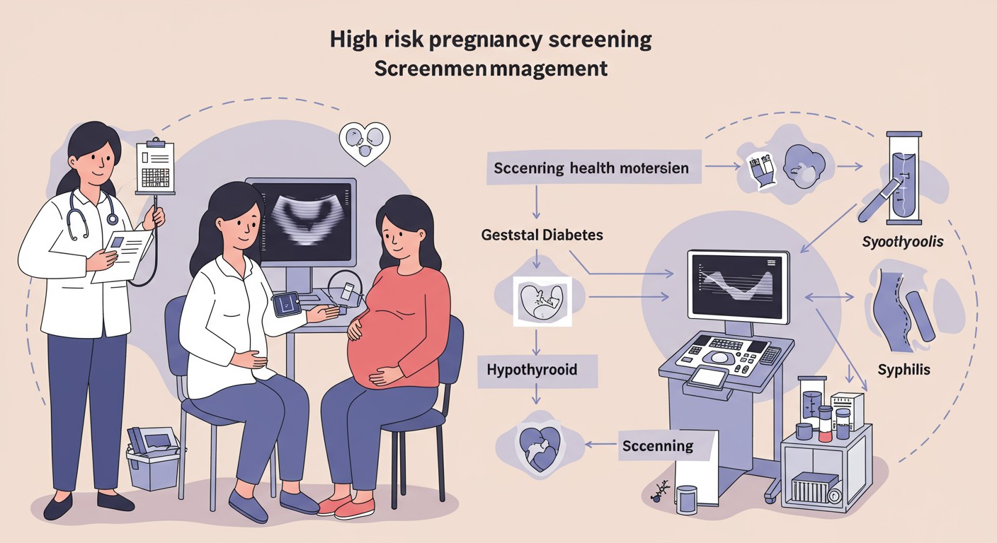
High-risk pregnancy screening process with community health nurse assessment and referral pathway
Core Components of High-Risk Approach
- Risk Assessment: Systematic evaluation to identify factors that increase the risk of adverse outcomes
- Early Screening: Timely detection of high-risk conditions through appropriate screening tests
- Risk Classification: Categorizing pregnant women based on risk level to determine appropriate care
- Primary Management: Initial interventions provided at the community level
- Referral System: Streamlined process for transferring high-risk cases to higher levels of care
- Follow-up: Monitoring of high-risk pregnancies throughout gestation
- Documentation: Systematic record-keeping of all interventions and outcomes
Advantage: The high-risk approach concentrates resources on those who need them most, making it potentially cost-effective for resource-limited settings.
Limitation: The approach may miss complications that develop in pregnancies initially categorized as low-risk, highlighting the need for continuous monitoring of all pregnancies.
3. Screening Overview and Principles
Screening in high-risk pregnancy refers to the systematic application of tests or procedures to identify conditions that may adversely affect maternal or fetal health. Effective screening is a cornerstone of high-risk pregnancy management in community health nursing.
Principles of Effective Screening
Characteristics of Good Screening Tests
- High sensitivity and specificity
- Cost-effective and accessible
- Acceptable to the target population
- Simple to perform and interpret
- Minimal discomfort and risk
Timing of Screening
- First Visit: Complete history, physical examination, baseline investigations
- Regular Antenatal Visits: Ongoing monitoring based on risk profile
- Specific Gestational Ages: Targeted screening for conditions like GDM (24-28 weeks)
Standard Screening Timeline
| Gestational Age | Standard Screening Tests | Additional Tests for High-Risk Cases |
|---|---|---|
| First Visit (<12 weeks) |
|
|
| 16-20 weeks |
|
|
| 24-28 weeks |
|
|
| 32-34 weeks |
|
|
| 36 weeks onwards |
|
|
4. High-Risk Complications: Screening and Management
4.1 Antepartum Hemorrhage
Antepartum hemorrhage (APH) is defined as bleeding from or into the genital tract occurring from 24 weeks of gestation until the birth of the baby. It complicates 2-5% of pregnancies and is a significant cause of perinatal and maternal morbidity and mortality.
Types and Causes
| Type | Definition | Risk Factors |
|---|---|---|
| Placenta Previa | Placenta implanted partially or completely over the internal cervical os |
|
| Placental Abruption | Premature separation of normally implanted placenta |
|
| Vasa Previa | Fetal vessels crossing the internal os without placental tissue |
|
| Other Causes | Cervical lesions, uterine rupture, marginal sinus bleeding |
|
Screening and Early Identification
Universal Screening
- Detailed obstetric history at first antenatal visit
- Second-trimester ultrasound (18-20 weeks) for placental location
- Follow-up scan at 32 weeks if low-lying placenta detected
High-Risk Specific Screening
- Additional ultrasound assessments for women with previous cesarean delivery
- Transvaginal ultrasound for precise placental localization
- Doppler studies to identify vasa previa in high-risk cases
Primary Management by Community Health Nurses
REMEMBER: Any APH is potentially life-threatening and requires immediate attention. Significant bleeding or hemodynamic instability constitutes an obstetric emergency.
- Initial Assessment
- Evaluate amount and nature of bleeding
- Assess vital signs (BP, pulse, respiratory rate)
- Check fetal heart sounds/rate
- Assess for uterine tenderness/contractions (without vaginal examination)
- Immediate Actions
- Establish IV access with large-bore cannula (16-18G)
- Start fluid resuscitation if signs of shock
- Position patient in left lateral position
- Administer oxygen if distressed
- DO NOT perform vaginal examination in suspected placenta previa
- Documentation
- Record time of onset, amount, color, and associated symptoms
- Document all vital signs and interventions
- Complete referral forms with all relevant information
B – Blood pressure and pulse monitoring
L – Left lateral position
E – Establish IV access
E – Evaluate fetal status
D – Dispatch to hospital immediately
Referral Criteria for APH
IMMEDIATE REFERRAL for ANY of the following:
- Any visible vaginal bleeding after 24 weeks
- Suspected placenta previa (painless bleeding)
- Suspected placental abruption (painful bleeding with uterine tenderness)
- Hemodynamic instability (tachycardia, hypotension)
- Abnormal fetal heart patterns
- Uterine contractions with bleeding
4.2 Pre-eclampsia and Eclampsia
Pre-eclampsia is a multisystem disorder specific to pregnancy characterized by hypertension and proteinuria after 20 weeks of gestation. Eclampsia refers to the occurrence of one or more convulsions in association with pre-eclampsia.
Definitions and Classification
| Condition | Definition | Diagnostic Criteria |
|---|---|---|
| Gestational Hypertension | New-onset hypertension after 20 weeks without proteinuria | BP ≥140/90 mmHg on two occasions at least 4 hours apart |
| Pre-eclampsia | Hypertension with evidence of end-organ damage |
|
| Severe Pre-eclampsia | Pre-eclampsia with severe features |
|
| Eclampsia | Convulsions in a woman with pre-eclampsia | Seizures that cannot be attributed to other causes |
| HELLP Syndrome | Variant of severe pre-eclampsia | Hemolysis, Elevated Liver enzymes, Low Platelets |
Risk Factors
Major Risk Factors
- Previous pre-eclampsia
- Chronic hypertension
- Pre-existing diabetes
- Renal disease
- Autoimmune disorders (SLE, antiphospholipid syndrome)
- Multiple pregnancy
Moderate Risk Factors
- First pregnancy
- Age ≥40 years
- BMI ≥35 kg/m²
- Family history of pre-eclampsia
- Pregnancy interval >10 years
- Previous adverse pregnancy outcome
Screening and Early Identification
First Trimester Screening
- Detailed history to identify risk factors
- Baseline BP measurement
- Urine protein assessment
- Combined screening with maternal factors and biomarkers (PAPP-A, PlGF) where available
Second Trimester Screening
- BP monitoring at each antenatal visit
- Urine dipstick for protein
- Uterine artery Doppler (20-24 weeks) in high-risk women
- Assessment for early signs of pre-eclampsia
Third Trimester Screening
- Increased frequency of BP monitoring
- Assessment of fetal growth and well-being
- Vigilance for symptoms of pre-eclampsia
- PlGF/sFlt-1 ratio testing where available
Warning Signs and Symptoms
S – Severe headache (unrelieved by acetaminophen)
C – Changes in vision (blurring, flashing lights, spots)
O – Oliguria (reduced urine output)
P – Pain in the upper right abdomen/epigastrium
E – Edema (sudden swelling of face, hands, or feet)
Primary Management by Community Health Nurses
- Regular Monitoring
- BP measurement at every antenatal visit
- Urine dipstick for protein
- Weight measurement to detect sudden gain
- Clinical assessment for edema
- Education on Warning Signs
- Educate about SCOPE warning signs
- Provide clear instructions on when to seek immediate medical attention
- Ensure understanding of the seriousness of pre-eclampsia
- Preventive Measures for High-Risk Women
- Low-dose aspirin (75-150 mg daily) starting at 12-16 weeks for high-risk women
- Calcium supplementation (1.5-2.0 g daily) in areas with low dietary calcium intake
- Regular monitoring of fetal growth and well-being
CAUTION: There is no single reliable, cost-effective screening test for pre-eclampsia. Vigilant clinical monitoring remains essential.
Management of Mild Pre-eclampsia at Community Level
- More frequent antenatal visits (at least weekly)
- Daily home BP monitoring if feasible
- Reduced physical activity with adequate rest
- Normal diet (no salt restriction)
- Education about danger signs requiring immediate attention
Emergency Management of Severe Pre-eclampsia/Eclampsia
Key Principle: Magnesium sulfate is the drug of choice for preventing and treating eclamptic seizures.
- Initial Stabilization (while arranging transfer)
- Position in left lateral position
- Secure airway and give oxygen if available
- Establish IV access
- If convulsing, protect from injury
- Magnesium Sulfate Administration (if available and authorized)
- Loading dose: 4g IV over 15-20 minutes
- Maintenance: 1g/hour by IV infusion
- Monitor for magnesium toxicity (respiratory rate, reflexes, urine output)
- Blood Pressure Control (if BP ≥160/110 mmHg)
- Labetalol 20mg IV slowly, then 40mg if needed after 10 minutes, then 80mg every 10 minutes to a maximum of 220mg
- OR Hydralazine 5-10mg IV every 20 minutes to a maximum of 20mg
- OR Nifedipine 10mg orally, repeated after 30 minutes if necessary
Referral Criteria
IMMEDIATE REFERRAL for:
- BP ≥140/90 mmHg with proteinuria
- BP ≥160/110 mmHg with or without proteinuria
- Any warning signs of pre-eclampsia (SCOPE)
- Proteinuria ≥1+ on dipstick
- Reduced fetal movements or abnormal fetal growth
- Eclamptic seizure (emergency transfer with magnesium sulfate if available)
4.3 Anemia in Pregnancy
Anemia in pregnancy is defined as hemoglobin (Hb) concentration below 11g/dL in the first and third trimesters, or below 10.5g/dL in the second trimester. It affects approximately 40% of pregnant women globally, with iron deficiency being the most common cause.
Classification of Anemia in Pregnancy
| Severity | Hemoglobin Level | Clinical Manifestations |
|---|---|---|
| Mild | 10.0-10.9 g/dL | Often asymptomatic or mild fatigue |
| Moderate | 7.0-9.9 g/dL | Fatigue, pallor, tachycardia, dyspnea on exertion |
| Severe | <7.0 g/dL | Marked fatigue, tachycardia, dyspnea at rest, increased risk of cardiac failure |
| Very Severe | <4.0 g/dL | Heart failure, tissue hypoxia, increased risk of maternal and fetal mortality |
Types and Causes of Anemia in Pregnancy
Common Types
- Iron Deficiency Anemia – Most common (>50% of cases)
- Folate Deficiency Anemia – Common in malnutrition
- Vitamin B12 Deficiency – Less common but serious
- Physiological Anemia of Pregnancy – Due to plasma volume expansion
Other Types
- Hemoglobinopathies – Sickle cell disease, thalassemia
- Anemia of Chronic Disease – Associated with infections, inflammation
- Aplastic Anemia – Rare but severe
- Hemolytic Anemia – Due to increased destruction of RBCs
Risk Factors
- Poor nutrition and dietary habits
- Multiple pregnancy
- Short interpregnancy interval (<2 years)
- Grand multiparity (≥5 pregnancies)
- Adolescent pregnancy
- Low socioeconomic status
- Parasitic infections (hookworm, malaria)
- Chronic conditions (inflammatory bowel disease, renal disease)
Screening and Diagnosis
Routine Screening
- Complete blood count (CBC) at first antenatal visit
- Repeat CBC at 24-28 weeks gestation
- Additional testing at 36 weeks or as needed
Additional Diagnostic Tests
- Serum ferritin (for iron deficiency)
- Peripheral blood smear
- Serum B12 and folate levels
- Hemoglobin electrophoresis (if hemoglobinopathy suspected)
Clinical pearl: Serum ferritin is the most sensitive test for iron deficiency, with levels <30 ng/mL indicating depleted iron stores even before anemia develops.
Primary Management by Community Health Nurses
I – Identify cause through appropriate screening
R – Recommend appropriate supplements
O – Optimize dietary intake of iron, folate, and vitamin B12
N – Note response to treatment and adjust accordingly
- Prevention and Prophylaxis
- Universal iron supplementation: 30-60 mg elemental iron daily
- Folic acid supplementation: 400 mcg daily (ideally started preconception)
- Nutritional counseling to increase dietary iron intake
- Deworming in endemic areas
- Treatment of Iron Deficiency Anemia
- Mild to moderate anemia (Hb 7-10.9 g/dL):
- Oral iron therapy: 120 mg elemental iron daily in 2-3 divided doses
- Continue for 3 months after hemoglobin normalization to replenish stores
- Medication adherence counseling:
- Take iron supplements with vitamin C sources to enhance absorption
- Avoid taking with calcium supplements, tea, coffee, or antacids
- Manage side effects (constipation, nausea, epigastric discomfort)
- Mild to moderate anemia (Hb 7-10.9 g/dL):
- Monitoring Response to Treatment
- Check hemoglobin levels 2-4 weeks after initiating treatment
- Expected increase: 1 g/dL within 2 weeks, 2 g/dL within 4 weeks
- Reassess if inadequate response
Referral Criteria
REFER to higher level of care for:
- Severe anemia (Hb <7 g/dL)
- Anemia with cardiac decompensation or respiratory distress
- Anemia not responding to oral iron therapy after 4 weeks
- Suspected hemoglobinopathy or other complex causes
- Anemia diagnosed late in pregnancy (>36 weeks)
- Iron intolerance requiring parenteral therapy
4.4 Gestational Diabetes Mellitus (GDM)
Gestational Diabetes Mellitus (GDM) is defined as glucose intolerance of variable severity with onset or first recognition during pregnancy. It affects approximately 2-10% of pregnancies worldwide, with increasing prevalence due to rising maternal obesity and age.
Risk Factors for GDM
Major Risk Factors
- Previous GDM
- Previous macrosomic baby (>4000g)
- BMI >30 kg/m²
- First-degree relative with diabetes
- Maternal age >35 years
- Previous unexplained stillbirth
Other Risk Factors
- Polycystic ovary syndrome
- Multiple pregnancy
- Ethnic groups with high prevalence (South Asian, Middle Eastern, African, Hispanic)
- Excessive gestational weight gain
- Current glycosuria
Screening and Diagnosis
Two main approaches exist for GDM screening:
| Approach | Method | Diagnostic Criteria |
|---|---|---|
| One-Step Approach (IADPSG/WHO) |
|
GDM diagnosed if any one value equals or exceeds:
|
| Two-Step Approach (ACOG) |
Step 1:
Step 2 (if Step 1 positive):
|
Step 1: Positive if 1-hour glucose ≥140 mg/dL (7.8 mmol/L) Step 2: GDM diagnosed if two or more values equal or exceed:
|
Early Screening: Women with high-risk factors should be screened for pre-existing diabetes at the first antenatal visit using standard diagnostic criteria. If negative, they should be rescreened at 24-28 weeks.
Primary Management by Community Health Nurses
- Initial Education and Counseling
- Explain GDM and its implications for mother and baby
- Emphasize the importance of glycemic control
- Assure that GDM usually resolves after delivery
- Inform about increased risk of type 2 diabetes in the future
- Lifestyle Modifications
- Medical Nutrition Therapy:
- Individualized diet plan (often 1,800-2,200 kcal/day)
- Carbohydrate distribution (40-45% of total calories)
- Complex carbohydrates instead of simple sugars
- Three meals and 2-3 snacks daily
- Physical Activity:
- Moderate-intensity exercise for 30 minutes daily
- Walking, swimming, or stationary cycling
- Avoid exercise with risk of falls or abdominal trauma
- Medical Nutrition Therapy:
- Blood Glucose Monitoring
- Self-monitoring of blood glucose (SMBG) 4 times daily:
- Fasting
- 1 or 2 hours after each main meal
- Target blood glucose levels:
- Fasting: ≤95 mg/dL (5.3 mmol/L)
- 1-hour postprandial: ≤140 mg/dL (7.8 mmol/L)
- 2-hour postprandial: ≤120 mg/dL (6.7 mmol/L)
- Self-monitoring of blood glucose (SMBG) 4 times daily:
S – Screening at appropriate time (24-28 weeks)
U – Understand blood glucose targets
G – Guide on diet and exercise
A – Assess need for insulin/medication
R – Regular monitoring of maternal and fetal wellbeing
Monitoring and Follow-up
- More frequent antenatal visits (every 2 weeks initially, then weekly after 36 weeks)
- Serial ultrasounds for fetal growth at 28, 32, and 36 weeks
- Monitoring for complications:
- Pre-eclampsia
- Polyhydramnios
- Fetal macrosomia
- Fetal surveillance from 32 weeks (if indicated):
- Non-stress test
- Biophysical profile
- Doppler studies
Referral Criteria
REFER to specialist care for:
- Diagnosis of GDM for initial assessment and management plan
- Inadequate glycemic control with lifestyle modifications alone
- Need for pharmacological therapy (insulin or oral agents)
- Evidence of fetal macrosomia or growth restriction
- Associated hypertension or other complications
- Difficulty in self-monitoring of blood glucose
Postpartum Follow-up
- Discontinue glucose-lowering medication immediately after delivery
- Screen for persistent diabetes at 6-12 weeks postpartum using 75g OGTT
- Annual screening for diabetes with fasting plasma glucose or HbA1c
- Counseling about:
- Risk reduction strategies for type 2 diabetes
- Preconception counseling for future pregnancies
- Contraception
4.5 Hypothyroidism in Pregnancy
Hypothyroidism in pregnancy is defined as an inadequate thyroid hormone production during pregnancy. It affects approximately 2-3% of pregnant women and can have significant adverse effects on maternal and fetal outcomes if untreated.
Types of Hypothyroidism in Pregnancy
| Type | Definition | Laboratory Findings |
|---|---|---|
| Overt Hypothyroidism | Clinical hypothyroidism with elevated TSH and reduced free T4 |
|
| Subclinical Hypothyroidism | Asymptomatic with elevated TSH but normal free T4 |
|
| Isolated Hypothyroxinemia | Normal TSH with low free T4 |
|
Important note: Pregnancy-specific reference ranges for TSH are lower than non-pregnant ranges:
- First trimester: 0.1-2.5 mIU/L
- Second trimester: 0.2-3.0 mIU/L
- Third trimester: 0.3-3.0 mIU/L
Risk Factors
- Personal history of thyroid disease
- Family history of thyroid disorders
- Presence of goiter
- Thyroid autoantibodies (TPOAb, TgAb)
- Previous radiation to head or neck
- Type 1 diabetes or other autoimmune disorders
- History of recurrent miscarriage or preterm delivery
- Age >30 years
- Iodine deficiency or excess
Screening and Diagnosis
Universal vs. Targeted Screening
There is ongoing debate about universal versus targeted screening for thyroid dysfunction in pregnancy:
- American Thyroid Association (ATA): Recommends targeted screening of high-risk women
- Some professional organizations: Support universal screening in the first trimester
Recommended Screening Tests
- TSH as the initial screening test
- If TSH is abnormal, measure Free T4
- In high-risk women, consider testing for thyroid peroxidase antibodies (TPOAb)
Effects of Untreated Hypothyroidism in Pregnancy
Maternal Effects
- Miscarriage
- Anemia
- Preeclampsia
- Placental abruption
- Postpartum hemorrhage
- Cardiac dysfunction
Fetal/Neonatal Effects
- Impaired neurocognitive development
- Low birth weight
- Preterm birth
- Increased perinatal mortality
- Congenital anomalies
Primary Management by Community Health Nurses
- Screening and Initial Assessment
- Targeted screening of high-risk women at first antenatal visit
- Assessment of signs and symptoms of hypothyroidism
- Referral for thyroid function tests when indicated
- Management of Women with Pre-existing Hypothyroidism
- Increase levothyroxine dose by approximately 30-50% as soon as pregnancy is confirmed
- Typically, this means adding two extra tablets per week
- Check TSH every 4-6 weeks during the first trimester, then once each in the second and third trimesters
- Adjust dose to maintain TSH within pregnancy-specific reference ranges
- Patient Education
- Take levothyroxine on an empty stomach (30-60 minutes before breakfast)
- Avoid taking with calcium, iron supplements, or antacids (separate by at least 4 hours)
- Importance of adherence to medication
- Need for increased dose during pregnancy
- Return to pre-pregnancy dose after delivery
T – Target screening in high-risk women
H – Higher dose requirements during pregnancy
Y – Yield better outcomes with early treatment
R – Regular monitoring of thyroid function
O – Optimize timing of medication intake
I – Iodine supplementation (150-250 μg daily)
D – Dose adjustment based on TSH levels
Treatment Guidelines
- Overt Hypothyroidism
- Immediate treatment with levothyroxine
- Starting dose: 1.6 μg/kg/day or 100-150 μg/day
- Higher starting dose for women with severe hypothyroidism
- Subclinical Hypothyroidism
- Treatment recommended if:
- TSH >10.0 mIU/L (regardless of TPOAb status)
- TSH >4.0 mIU/L with positive TPOAb
- TSH >2.5 mIU/L with symptoms of hypothyroidism
- Starting dose: 50-75 μg/day
- Treatment recommended if:
- Isolated Hypothyroxinemia
- Insufficient evidence for treatment
- Consider treatment in areas with known iodine deficiency
Referral Criteria
REFER to specialist care for:
- Newly diagnosed overt hypothyroidism in pregnancy
- Difficulty achieving target TSH levels despite treatment
- Pre-existing hypothyroidism with unstable control
- Thyroid nodules or goiter
- TPOAb positivity with normal thyroid function
- Complex thyroid disorders (e.g., post-thyroidectomy, history of thyroid cancer)
Postpartum Considerations
- Reduce levothyroxine to pre-pregnancy dose immediately after delivery
- Check TSH at 6 weeks postpartum and adjust dose as needed
- Monitor for postpartum thyroiditis, especially in TPOAb-positive women
- Levothyroxine is safe during breastfeeding
4.6 Syphilis in Pregnancy
Syphilis is a sexually transmitted infection caused by the spirochete Treponema pallidum. When acquired during pregnancy, it can lead to severe adverse outcomes including congenital syphilis, which can cause stillbirth, neonatal death, prematurity, and long-term sequelae in surviving infants.
Stages of Syphilis
| Stage | Timeframe | Clinical Manifestations | Infectivity |
|---|---|---|---|
| Primary | 3-90 days after exposure | Painless chancre at site of inoculation | Highly infectious |
| Secondary | 2-12 weeks after primary stage | Generalized rash (often palms/soles), mucocutaneous lesions, lymphadenopathy, fever, malaise | Highly infectious |
| Latent Early | <1 year after infection | Asymptomatic | Potentially infectious |
| Latent Late | >1 year after infection | Asymptomatic | Minimally infectious except to fetus |
| Tertiary | 10-30 years after infection | Gummatous lesions, cardiovascular and neurological manifestations | Not infectious except to fetus |
Risk Factors for Syphilis in Pregnancy
- Multiple sexual partners
- Unprotected sexual intercourse
- Other sexually transmitted infections
- Substance abuse
- Late or inadequate prenatal care
- Young age (<25 years)
- Commercial sex work
- Residence in high-prevalence areas
Screening and Diagnosis
Screening Recommendations
- Universal screening for all pregnant women at:
- First prenatal visit (mandatory in most countries)
- Third trimester (28-32 weeks), especially in high-risk women
- Delivery for women with inadequate prenatal care or in high-prevalence areas
- Additional screening after exposure to an infected partner
Diagnostic Approach
Traditional Algorithm:
- Initial non-treponemal test (RPR or VDRL)
- If positive, confirm with treponemal test (TP-PA, FTA-ABS, etc.)
Reverse Algorithm:
- Initial treponemal test (EIA or CIA)
- If positive, perform quantitative non-treponemal test (RPR or VDRL)
Clinical pearl: Non-treponemal tests (RPR, VDRL) measure disease activity and can be used to monitor treatment response. Treponemal tests remain positive for life once infected, regardless of treatment.
Effects of Untreated Syphilis in Pregnancy
Untreated maternal syphilis can lead to congenital syphilis in up to 80% of cases, with outcomes including:
- Spontaneous abortion (40% risk in early syphilis)
- Stillbirth
- Hydrops fetalis
- Preterm birth
- Low birth weight
- Neonatal death
- Early congenital syphilis (first 2 years of life): hepatosplenomegaly, snuffles, rash, pseudoparalysis
- Late congenital syphilis: Hutchinson’s teeth, mulberry molars, saddle nose, saber shins, neurological sequelae
Primary Management by Community Health Nurses
S – Screening at first prenatal visit
Y – Yield better outcomes with early detection
P – Penicillin is the only effective treatment
H – High-risk women need repeat screening
I – Involve partners in testing and treatment
L – Laboratory monitoring after treatment
I – Immediate treatment prevents congenital transmission
S – Secondary prevention through safe sex education
- Screening Implementation
- Ensure universal screening at first antenatal visit
- Implement systems to ensure test results are received and acted upon
- Provide repeat screening for high-risk women in the third trimester
- Treatment
- Penicillin G is the only proven effective treatment for preventing maternal transmission and treating fetal infection
- Treatment regimen based on stage:
- Early syphilis (primary, secondary, early latent): Benzathine penicillin G 2.4 million units IM single dose
- Late latent or unknown duration: Benzathine penicillin G 2.4 million units IM weekly for 3 weeks
- For penicillin-allergic patients: Desensitization and penicillin treatment is recommended (alternative antibiotics are not proven effective for fetal treatment)
- Partner Notification and Treatment
- Identify and notify partners for testing and treatment
- Maintain confidentiality while ensuring public health protection
- Educate about safe sex practices
- Jarisch-Herxheimer Reaction Management
- Educate patients about this possible reaction (fever, chills, headache, myalgia occurring within 24 hours of treatment)
- Provide symptomatic management with antipyretics
- Monitor fetal status in pregnant women as it may induce preterm labor
- Follow-up Monitoring
- Clinical assessment at 1 month after treatment
- Serological monitoring with non-treponemal tests at 1, 3, 6, and 12 months
- Adequate response: Four-fold decrease in non-treponemal titer within 6-12 months
Referral Criteria
REFER to specialist care for:
- Confirmed syphilis diagnosis for appropriate staging and treatment
- Penicillin allergy requiring desensitization
- Suspected tertiary syphilis or neurosyphilis
- HIV co-infection
- Failed treatment (persistent symptoms or inadequate serological response)
- Treatment in third trimester (high risk of congenital syphilis)
5. Referral System and Criteria
An effective referral system is crucial for the successful implementation of the high-risk approach in pregnancy. It ensures that women with complications receive timely and appropriate care at facilities with adequate resources.
Components of an Effective Referral System
Identification
- Clear criteria for referral
- Standardized screening tools
- Training of frontline workers
- Defined pathways based on risk levels
Communication
- Standardized referral forms
- Direct communication channels
- Feedback mechanisms
- Clear documentation
Logistics
- Transportation arrangements
- Emergency protocols
- Financial considerations
- Facility readiness
Levels of Care in Referral System
| Level | Facility Type | Services Available | Staffing |
|---|---|---|---|
| Primary | Health centers, Community clinics, Sub-centers |
|
|
| Secondary | District hospitals, First referral units |
|
|
| Tertiary | Regional/teaching hospitals, Specialized centers |
|
|
Standardized Referral Documentation
A comprehensive referral form should include:
- Patient Information: Name, age, contact details, unique ID
- Obstetric History: Gravidity, parity, LMP, EDD, gestational age
- Current Pregnancy Details: Risk factors, complications
- Vital Signs: BP, pulse, temperature, respiratory rate
- Examination Findings: General, systemic, obstetric
- Laboratory Results: Relevant investigations
- Interventions Already Provided: Treatments, procedures
- Reason for Referral: Specific indication
- Urgency Level: Emergency, urgent, routine
- Referring Provider Details: Name, designation, contact information
Referral Decision Pathway
- Assess woman at antenatal visit or when complication arises
- Identify risk factors or complications using standardized screening tools
- Determine if condition can be managed at current level
- If YES → Provide appropriate care and follow-up
- If NO → Proceed to referral process
- Classify urgency of referral
- Emergency (immediate): Life-threatening conditions
- Urgent (same day): Conditions requiring prompt attention
- Semi-urgent (within 72 hours): Conditions requiring early assessment
- Routine (within 1-2 weeks): Non-urgent conditions requiring higher-level care
- Stabilize patient before transfer if emergency situation
- Communicate with receiving facility
- Arrange transportation as needed
- Complete referral documentation
- Provide clear instructions to the woman and family
- Follow up to ensure referral was completed and feedback received
Common Barriers to Effective Referral
System-Related Barriers
- Poor communication between referral levels
- Lack of standardized protocols
- Inadequate transportation
- Limited capacity at referral facilities
- Insufficient feedback mechanisms
Patient-Related Barriers
- Financial constraints
- Distance to referral facility
- Cultural and social factors
- Lack of understanding about severity
- Fear of unfamiliar environment
Strategies to Improve Referral System
- Standardization: Develop and implement clear referral protocols and forms
- Training: Ensure healthcare providers at all levels understand referral criteria
- Communication: Establish reliable communication channels between facilities
- Transportation: Develop emergency transport strategies for obstetric emergencies
- Feedback: Create mechanisms for referral feedback and continuous improvement
- Community Engagement: Educate communities about the importance of referrals
- Monitoring: Track referral patterns and outcomes to identify system gaps
6. Follow-up Protocols
Follow-up care is an essential component of the high-risk approach to ensure continuity of care and monitor the progression of high-risk conditions. Proper follow-up protocols help prevent complications, adjust management plans, and improve overall maternal and fetal outcomes.
General Principles of Follow-up
- Individualization: Tailor frequency and content based on specific risk factors
- Communication: Ensure seamless information sharing between different care providers
- Compliance monitoring: Track adherence to treatment and recommendations
- Reassessment: Continuously evaluate for new or worsening risk factors
- Proactive surveillance: Anticipate potential complications based on risk profile
- Documentation: Maintain detailed records of all follow-up visits and findings
Follow-up Schedule by Risk Category
| Risk Category | Recommended Follow-up Schedule | Assessment Components |
|---|---|---|
| Low Risk |
|
|
| Moderate Risk |
|
|
| High Risk |
|
|
Condition-Specific Follow-up Guidelines
Antepartum Hemorrhage
- After initial stabilization/management:
- Weekly antenatal visits if placenta previa with no active bleeding
- Serial ultrasounds every 2-3 weeks to assess placental location and fetal growth
- Admission to hospital for any recurrent bleeding
- Monitoring for anemia and correcting as needed
- Hospitalization at 34-36 weeks for high-risk cases
- Planned delivery timing based on clinical status and placental location
Pre-eclampsia
- Mild pre-eclampsia:
- Twice weekly BP checks (home or facility)
- Weekly urine protein assessment
- Laboratory tests (CBC, liver and renal function) every 1-2 weeks
- Fetal growth assessment every 3 weeks
- NST and AFI assessment weekly after 32 weeks
- Daily fetal movement counting
- Severe pre-eclampsia (post-hospitalization):
- More intensive monitoring or continued hospitalization
- Daily BP monitoring
- Twice weekly laboratory tests
- NST 2-3 times per week
- Weekly Doppler assessment
- Timing delivery based on maternal and fetal status
Anemia
- Mild to moderate anemia:
- Hemoglobin check 2-4 weeks after starting treatment
- Monthly hemoglobin assessment until normalized
- Evaluate compliance with iron therapy
- Assess for side effects of iron supplementation
- Continue iron for 3 months after hemoglobin normalization
- Severe anemia (after initial treatment):
- Weekly hemoglobin checks until levels >7g/dL
- Then follow moderate anemia protocol
- Assess for underlying causes if poor response to treatment
Gestational Diabetes
- Diet-controlled GDM:
- Fortnightly antenatal visits
- Blood glucose self-monitoring 4 times daily
- Weekly review of glucose logs
- Monthly HbA1c
- Growth scans at 28, 32, and 36 weeks
- Weekly NST after 36 weeks
- Medication-treated GDM:
- Weekly antenatal visits
- Twice weekly reviews of glucose logs
- Medication adjustments as needed
- Monthly HbA1c
- Growth scans every 3-4 weeks
- Twice weekly NST after 36 weeks
- Postpartum follow-up:
- 75g OGTT at 6-12 weeks postpartum
- Annual screening for diabetes
Hypothyroidism
- Pre-existing hypothyroidism:
- TSH every 4-6 weeks in first trimester
- TSH once in second trimester
- TSH once in third trimester
- Adjust levothyroxine dose based on TSH levels
- Newly diagnosed hypothyroidism:
- TSH 4 weeks after initiating treatment
- Then follow protocol for pre-existing hypothyroidism
- Assess for improvement in any symptoms
- Postpartum follow-up:
- Reduce to pre-pregnancy dose immediately after delivery
- TSH at 6 weeks postpartum
- Annual thyroid function testing
Syphilis
- After treatment:
- Clinical assessment at 1 month
- Quantitative non-treponemal tests (RPR/VDRL) at 1, 3, 6, and 12 months
- Expected 4-fold decline in titers within 6-12 months
- Re-treatment if signs/symptoms persist or titers increase by 4-fold
- Ensure partner treatment and follow-up
- Late pregnancy diagnosis:
- More intensive surveillance
- Monthly serological monitoring until delivery
- Detailed planning for neonatal evaluation and treatment
Follow-up After Referral
- Communication with referral facility: Obtain detailed feedback about diagnosis, treatment, and follow-up plan
- Counter-referral documentation: Receive formal counter-referral form with clear instructions
- Continued care coordination: Clarify roles and responsibilities between primary and specialized care
- First follow-up visit: Schedule within timeframe recommended by referral facility
- Review treatment plan: Ensure understanding and compliance with recommended regimen
- Adjust follow-up schedule: Modify frequency based on current risk assessment
- Re-referral criteria: Define clear indicators for when to re-refer to specialized care
Patient Engagement in Follow-up
Strategies to Improve Compliance
- Reminder systems (text messages, phone calls)
- Appointment cards with clear dates and times
- Education about importance of follow-up
- Family involvement in care plan
- Community health worker home visits
- Flexible clinic hours to accommodate working women
- Combination of follow-up visits when possible
Self-Monitoring Components
- Home blood pressure monitoring
- Blood glucose self-testing
- Daily fetal movement counting
- Symptom diaries
- Medication compliance tracking
- Weight monitoring
- Recognition of warning signs requiring immediate attention
7. Record Maintenance and Reporting
Comprehensive and systematic documentation is a critical component of high-risk pregnancy management. Proper record maintenance facilitates continuity of care, enables effective communication between healthcare providers, supports clinical decision-making, and provides data for quality improvement and research.
Essential Components of Record Keeping
Patient Information
- Demographic details
- Contact information
- Emergency contacts
- Socioeconomic factors
- Educational status
- Occupation
- Health insurance information
Obstetric History
- Gravidity and parity
- Previous pregnancy outcomes
- Complications in prior pregnancies
- Mode of previous deliveries
- Birth weights
- Gestational age at delivery
- Interpregnancy interval
Current Pregnancy Details
- LMP and EDD
- Dating scan details
- Risk assessment findings
- Antenatal visit schedule
- Immunization status
- Supplementation details
- Birth plan
Documentation for High-Risk Conditions
| Condition | Essential Documentation Elements |
|---|---|
| Antepartum Hemorrhage |
|
| Pre-eclampsia/Eclampsia |
|
| Anemia |
|
| Gestational Diabetes |
|
| Hypothyroidism |
|
| Syphilis |
|
Referral Documentation
Outgoing Referral Record
- Date and time of referral
- Reason for referral
- Urgency level
- Clinical summary
- Investigations done
- Treatments provided
- Provisional diagnosis
- Mode of transportation
- Accompanying healthcare provider (if any)
- Receiving facility and provider
- Information provided to patient/family
Incoming Referral/Counter-referral
- Date of assessment at referral facility
- Confirmed diagnosis
- Treatment provided
- Current medications
- Follow-up plan
- Next appointment at referral facility
- Instructions for primary care provider
- Warning signs requiring re-referral
- Contact information for queries
Record Formats
Paper-Based Records
- Maternal Health Card: Carried by the woman
- Facility Register: Maintained at the health facility
- High-Risk Register: Specific for high-risk cases
- Referral Forms: For communication between facilities
- Laboratory Registers: For test results
- Follow-up Cards: For tracking scheduled visits
Electronic Health Records
- Integrated systems linking all levels of care
- Decision support tools for high-risk conditions
- Automated reminders for follow-up appointments
- Data analytics for monitoring outcomes
- Telemedicine platforms for remote consultations
- Mobile health applications for patient engagement
Reporting Systems
- Routine Health Information Systems (RHIS)
- Monthly/quarterly aggregated reports
- Indicators on antenatal coverage, high-risk detection, referrals
- Facility-based outcome metrics
- Maternal and Perinatal Death Surveillance and Response (MPDSR)
- Notification of all maternal and perinatal deaths
- Review process to identify contributing factors
- Response mechanisms to address identified issues
- Special Surveillance Systems
- Congenital syphilis surveillance
- Gestational diabetes registry
- Pre-eclampsia monitoring systems
- High-risk pregnancy outcome tracking
Key Performance Indicators for Monitoring
| Category | Indicators |
|---|---|
| Coverage |
|
| Quality |
|
| Outcome |
|
| Process |
|
Data Quality and Confidentiality
- Data Quality Assurance
- Regular audits of records for completeness and accuracy
- Training on proper documentation
- Data validation processes
- Standardized documentation formats
- Confidentiality and Privacy
- Secure storage of physical records
- Password protection for electronic records
- Limited access to authorized personnel
- De-identification of data for reporting purposes
- Informed consent for sharing information
8. Mnemonics and Learning Aids
Mnemonics are valuable memory tools that can help nursing students remember complex information related to high-risk pregnancy screening, management, and follow-up. Here’s a compilation of useful mnemonics for quick reference:
A – Age (extremes: <18 or >35 years)
B – Background medical conditions (diabetes, hypertension, thyroid disorders)
C – Complications in previous pregnancies
D – Delayed prenatal care or inadequate visits
E – Environmental/social factors (substance abuse, domestic violence)
S – Systematic approach to risk identification
C – Comprehensive history taking
R – Regular reassessment throughout pregnancy
E – Evidence-based screening tests
E – Effective documentation of findings
N – Nurturing therapeutic relationship with patient
S – Severe headache (unrelieved by acetaminophen)
C – Changes in vision (blurring, flashing lights, spots)
O – Oliguria (reduced urine output)
P – Pain in the upper right abdomen/epigastrium
E – Edema (sudden swelling of face, hands, or feet)
B – Blood pressure and pulse monitoring
L – Left lateral position
E – Establish IV access
E – Evaluate fetal status
D – Dispatch to hospital immediately
I – Identify cause through appropriate screening
R – Recommend appropriate supplements
O – Optimize dietary intake of iron, folate, and vitamin B12
N – Note response to treatment and adjust accordingly
S – Screening at appropriate time (24-28 weeks)
U – Understand blood glucose targets
G – Guide on diet and exercise
A – Assess need for insulin/medication
R – Regular monitoring of maternal and fetal wellbeing
T – Target screening in high-risk women
H – Higher dose requirements during pregnancy
Y – Yield better outcomes with early treatment
R – Regular monitoring of thyroid function
O – Optimize timing of medication intake
I – Iodine supplementation (150-250 μg daily)
D – Dose adjustment based on TSH levels
S – Screening at first prenatal visit
Y – Yield better outcomes with early detection
P – Penicillin is the only effective treatment
H – High-risk women need repeat screening
I – Involve partners in testing and treatment
L – Laboratory monitoring after treatment
I – Immediate treatment prevents congenital transmission
S – Secondary prevention through safe sex education