
Intrauterine Fetal Death (IUFD)
Comprehensive Notes for Nursing Students
Table of Contents
Introduction to Intrauterine Fetal Death
Definition
Intrauterine fetal death (IUFD) refers to the death of a fetus at or after 20 weeks of gestation or with a birth weight of at least 500 grams. It is characterized by the cessation of fetal cardiac activity, movement, and blood flow prior to complete expulsion from the mother.
The World Health Organization (WHO) defines intrauterine fetal death as “death prior to the complete expulsion or extraction from its mother of a product of conception, irrespective of the duration of pregnancy.” For statistical purposes, the WHO further specifies that IUFD generally refers to deaths of fetuses weighing 500g or more, or those that occurred after 22 completed weeks of gestation.
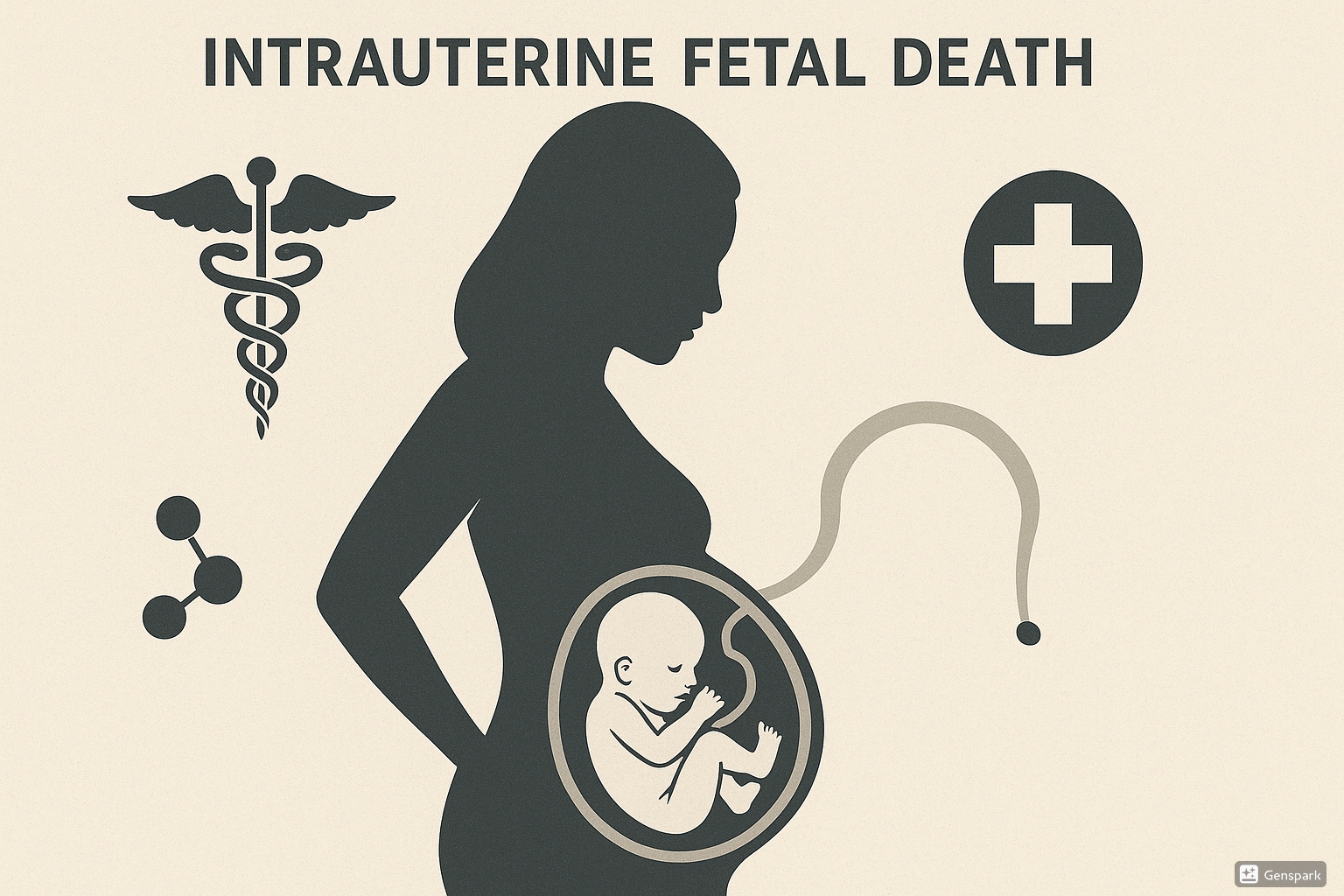
Medical illustration depicting intrauterine fetal death showing a silhouette of pregnant woman with fetus
Terminology Clarification
- Stillbirth: Often used interchangeably with IUFD, but specifically refers to the delivery of a fetus without signs of life.
- Fetal demise: Another term used to describe intrauterine fetal death.
- Miscarriage: Generally refers to pregnancy loss before 20 weeks of gestation.
Epidemiology
Understanding the prevalence and distribution of intrauterine fetal death is crucial for developing effective preventive strategies and improving maternal care.
| Region | IUFD Rate (per 1000 births) | Notable Factors |
|---|---|---|
| High-income countries | 3-5 | Advanced maternal age, obesity, smoking |
| Middle-income countries | 10-20 | Limited access to antenatal care, maternal infections |
| Low-income countries | 25-40 | Poor nutrition, inadequate healthcare infrastructure, untreated maternal conditions |
Key Epidemiological Facts
- Globally, an estimated 2.6 million cases of intrauterine fetal death occur annually
- 98% of cases occur in low and middle-income countries
- Approximately 1 in 160 pregnancies in the United States results in stillbirth
- The rate of unexplained IUFD has decreased with improvements in diagnostic capabilities
- Racial disparities exist, with higher rates observed among Black women compared to other racial groups
Etiology
The causes of intrauterine fetal death are diverse and often multifactorial. Understanding these causes is essential for prevention and management strategies.
Maternal Factors
- Advanced maternal age (>35 years)
- Hypertensive disorders (preeclampsia, eclampsia)
- Diabetes mellitus (pre-existing or gestational)
- Autoimmune disorders (antiphospholipid syndrome, SLE)
- Infections (malaria, syphilis, listeriosis, parvovirus B19)
- Thyroid disorders
- Substance abuse (tobacco, alcohol, drugs)
- Severe trauma
Placental Factors
- Placental abruption
- Placenta previa
- Placental insufficiency
- Vasa previa
- Umbilical cord accidents (prolapse, true knots)
- Velamentous cord insertion
- Chorioamnionitis
Fetal Factors
- Chromosomal abnormalities (trisomies)
- Congenital anomalies
- Intrauterine growth restriction (IUGR)
- Multiple gestation complications
- Hydrops fetalis
- Fetal-maternal hemorrhage
Mnemonic: “DEATH CAUSES”
- D: Diabetes and maternal Diseases
- E: Eclampsia and hypertensive disorders
- A: Abruption of placenta
- T: Trauma and Thrombophilias
- H: Hydrops and fetal anomalies
- C: Cord accidents
- A: Autoimmune disorders
- U: Uterine rupture
- S: Smoking and Substance abuse
- E: Extreme maternal age
- S: Sepsis and Infections
Risk Factors
Numerous risk factors have been associated with an increased likelihood of intrauterine fetal death. Recognizing these factors is essential for appropriate surveillance and preventive interventions.
| Category | Risk Factors | Relative Risk |
|---|---|---|
| Maternal Demographics |
|
1.5-3.0 |
| Obstetric History |
|
2.0-5.0 |
| Medical Conditions |
|
2.0-10.0 |
| Lifestyle Factors |
|
1.5-3.0 |
| Current Pregnancy |
|
2.0-4.0 |
High-Alert Risk Factors
The following risk factors are associated with particularly high risk of intrauterine fetal death and warrant enhanced surveillance:
- Previous stillbirth (3-4 fold increased risk)
- Uncontrolled diabetes (5-10 fold increased risk)
- Severe placental insufficiency (4-8 fold increased risk)
- Antiphospholipid syndrome (3-5 fold increased risk)
- Severe fetal growth restriction (4-8 fold increased risk)
Pathophysiology
The pathophysiological mechanisms leading to intrauterine fetal death are complex and often involve a sequence of events that ultimately result in fetal demise. Understanding these mechanisms is crucial for developing preventive strategies.
Common Pathophysiological Pathways
Placental Insufficiency
Reduction in placental function leading to:
- Decreased nutrient transfer
- Impaired oxygen exchange
- Accumulation of waste products
- Fetal hypoxia and acidosis
- Growth restriction
Hypoxic-Ischemic Injury
Progressive oxygen deprivation leading to:
- Myocardial dysfunction
- Multiorgan failure
- Metabolic acidosis
- Circulatory collapse
- Cardiac arrest
Infection/Inflammation
Infectious process causing:
- Direct fetal infection
- Inflammatory cytokine release
- Chorioamnionitis
- Placental dysfunction
- Preterm labor
Pathophysiological Sequence in IUFD
- Initial insult or abnormality
- Compromise of fetal-placental unit
- Progressive fetal hypoxemia
- Fetal compensatory mechanisms (blood redistribution to vital organs)
- Decompensation and multiorgan failure
- Cardiac arrest and intrauterine fetal death
- Post-mortem changes (maceration, autolysis)
Maceration Process After IUFD
After intrauterine fetal death occurs, predictable changes occur to the fetal body if retention in utero continues:
| Time After Death | Observable Changes |
|---|---|
| 0-8 hours | No maceration visible |
| 12-24 hours | Skin slippage, reddish discoloration of umbilical cord |
| 2-3 days | Skin vesicles form, extensive skin peeling, red-brown amniotic fluid |
| 4-7 days | Liver becoming darker, body cavities contain blood-stained fluid |
| 1-2 weeks | Extensive skin loss, skull bones become loose and overlapping |
| >2 weeks | Mummification may begin, organs difficult to identify |
Clinical Presentation
The clinical presentation of intrauterine fetal death can vary significantly. Understanding these signs and symptoms is crucial for early recognition and appropriate management.
Common Presenting Features
- Absence of fetal movements – Most common presenting symptom (80-90%)
- Absence of fetal heart tones on auscultation or Doppler examination
- Regression of pregnancy symptoms (nausea, breast tenderness)
- Vaginal bleeding or spotting (30-40% of cases)
- Sensation of “heaviness” in the abdomen
- Dark or brownish vaginal discharge
- Lack of expected fundal height growth
Important Clinical Pearls
- Many women with intrauterine fetal death may initially be asymptomatic
- Approximately 50% of women report a gradual decrease in fetal movements prior to complete cessation
- Some women may report a “premonition” that something is wrong with their pregnancy
- In cases of twin pregnancy, one fetus may be viable while the other has experienced IUFD
Maternal Psychological Response
The psychological response upon diagnosis of intrauterine fetal death may include:
- Acute grief reaction
- Denial and disbelief
- Guilt and self-blame
- Anger and resentment
- Fear and anxiety
- Emotional numbness
- Depression
- Physical manifestations of grief
- Uncertainty about the future
- Concerns about future pregnancies
Mnemonic: “FETAL DEMISE”
- F: Fetal movement cessation
- E: Empty feeling in abdomen
- T: Tissue discharge (brownish)
- A: Absence of fetal heart tones
- L: Loss of pregnancy symptoms
- D: Discharge (brownish or dark)
- E: Emotional distress/premonition
- M: Missed growth of fundal height
- I: Irregular uterine contractions
- S: Spotting or bleeding
- E: Edema reduction (if previously present)
Diagnosis
The diagnosis of intrauterine fetal death requires a systematic approach combining clinical assessment and confirmatory imaging studies. Early and accurate diagnosis is crucial for appropriate management.
Clinical Assessment
- Detailed history of maternal symptoms
- Absence of fetal movements
- Absence of fetal heart sounds on auscultation
- No fetal heart activity on handheld Doppler
- Assessment of fundal height (may be smaller than expected)
Ultrasonographic Findings
Definitive diagnostic criteria:
- Absence of cardiac activity (documented for at least 30 seconds)
- Absence of fetal movement during extended observation
- Spalding’s sign (overlapping skull bones)
- Collapse of fetal skull (if maceration present)
Additional Imaging Findings
- Robert’s sign (gas bubbles in fetal circulation)
- Hyperechogenic brain tissue
- Abnormal fetal posture
- Fetal edema or hydrops
- Oligohydramnios or anhydramnios
- Placental changes
Important Diagnostic Considerations
- Diagnosis of intrauterine fetal death must always be confirmed by ultrasound
- A second opinion should be sought whenever possible
- Documentation should include images confirming absence of cardiac activity
- In twin pregnancies, careful assessment of both fetuses is essential
Post-Diagnosis Investigations
After confirming intrauterine fetal death, the following investigations should be considered to identify the cause:
| Investigation Category | Specific Tests | Purpose |
|---|---|---|
| Maternal Blood Tests |
|
Identify maternal conditions and complications |
| Serological Tests |
|
Identify infectious or immunological causes |
| Post-Delivery Investigations |
|
Determine specific fetal or placental abnormalities |
Management
The management of intrauterine fetal death requires a multidisciplinary approach addressing medical, emotional, and psychological aspects of care. The goal is to safely deliver the fetus while providing comprehensive support to the mother and family.
Delivery Planning
Once intrauterine fetal death is confirmed, decisions regarding the timing and method of delivery must be made:
Expectant Management
Considerations:
- Appropriate for women who prefer to wait for spontaneous labor
- 85-90% of women will spontaneously labor within 2-3 weeks
- Requires close monitoring for complications
- Weekly coagulation studies to monitor for DIC
- Not recommended beyond 3-4 weeks due to risk of coagulopathy
Active Management
Methods of Induction:
- Medical induction with misoprostol or mifepristone-misoprostol combination
- Cervical ripening with prostaglandin E2
- Oxytocin augmentation if needed
- Mechanical methods (foley catheter, laminaria)
- Cesarean delivery rarely indicated except for specific maternal indications
Medical Induction Protocol
For intrauterine fetal death in the second or third trimester:
- Mifepristone: 200mg orally followed 24-48 hours later by misoprostol
- Misoprostol:
- 13-26 weeks: 100-200 μg vaginally every 4-6 hours
- 27-42 weeks: 25-50 μg vaginally every 4-6 hours
- Oxytocin: May be added if contractions inadequate after misoprostol
Management of Complications
| Complication | Signs/Symptoms | Management |
|---|---|---|
| Disseminated Intravascular Coagulation (DIC) | Abnormal bleeding, petechiae, ecchymosis, abnormal coagulation studies |
|
| Infection | Fever, uterine tenderness, foul-smelling discharge, elevated WBC |
|
| Psychological Trauma | Acute grief, depression, anxiety, post-traumatic stress |
|
| Retained Placental Tissue | Prolonged bleeding, incomplete delivery of placenta |
|
Pain Management
Adequate pain management is essential during delivery following intrauterine fetal death:
- Neuraxial analgesia (epidural) is recommended when available
- Patient-controlled analgesia for intravenous opioids
- Non-pharmacological methods (positioning, relaxation techniques)
- Consider sedation options if extreme emotional distress
Complications
Intrauterine fetal death can lead to various complications that require vigilant monitoring and prompt management.
Maternal Physical Complications
- Coagulopathy and DIC: Risk increases with retention time
- Infection: Chorioamnionitis, endometritis
- Labor complications: Prolonged labor, dystocia
- Postpartum hemorrhage: Due to uterine atony
- Sepsis: If infection progresses
Psychological Complications
- Complicated grief: Prolonged or intensified grief response
- Depression: Major depressive disorder may develop
- Anxiety disorders: Including PTSD
- Relationship difficulties: Strain on partnerships
- Fear about future pregnancies: Tokophobia
Long-term Impacts
- Increased risk in subsequent pregnancies: 2-10 fold higher risk of recurrence
- Chronic psychological sequelae: Anniversary reactions
- Impact on other children: Siblings may be affected
- Changes in family dynamics: Altered relationships
- Altered self-perception: Impact on maternal identity
Risk Factors for Psychological Complications
The following factors may increase the risk of developing significant psychological complications after intrauterine fetal death:
- Previous mental health conditions
- Poor social support
- Multiple pregnancy losses
- Traumatic delivery experience
- Perceived inadequate care during the event
- Pre-existing relationship difficulties
- History of trauma or adverse childhood experiences
Mnemonic: “COPE WITH LOSS”
Recognizing complications after intrauterine fetal death:
- C: Coagulopathy (DIC)
- O: Obstetric complications (prolonged labor)
- P: Psychological trauma (PTSD, depression)
- E: Endometritis and infection
- W: Weakened relationships
- I: Identity crisis
- T: Tokophobia (fear of future pregnancies)
- H: Hemorrhage risk
- L: Lactation issues
- O: Obsessive thoughts about loss
- S: Sepsis risk
- S: Subsequent pregnancy complications
Nursing Care
Nursing care for patients experiencing intrauterine fetal death requires a comprehensive approach that addresses both physical and emotional needs. The nurse plays a pivotal role in providing compassionate, patient-centered care during this traumatic experience.
Immediate Nursing Interventions
- Provide a private, quiet environment
- Allow family members to be present for support
- Use clear, compassionate communication
- Monitor vital signs and assess for complications
- Administer medications as ordered
- Collect laboratory specimens as needed
- Document findings accurately
Intrapartum Care
- Provide continuous emotional support
- Assist with pain management techniques
- Monitor progress of labor
- Assess for complications (hemorrhage, infection)
- Prepare for and assist with delivery
- Create mementos if desired (handprints, footprints)
- Facilitate cultural or religious practices
Postpartum Care
- Monitor for physical complications
- Provide lactation suppression information
- Assess for signs of grief and psychological distress
- Provide information about funeral arrangements
- Facilitate seeing and holding the baby if desired
- Provide information about support resources
- Arrange follow-up appointments
Nursing Care Plan for IUFD
| Nursing Diagnosis | Interventions | Expected Outcomes |
|---|---|---|
| Acute Grief related to loss of pregnancy as evidenced by crying, expressions of sorrow, and verbalization of loss |
|
|
| Risk for Complicated Grieving related to sudden loss and previous mental health issues |
|
|
| Risk for Infection related to tissue trauma and prolonged rupture of membranes |
|
|
| Risk for Deficient Fluid Volume related to potential hemorrhage |
|
|
Documentation Guidelines
Comprehensive documentation for intrauterine fetal death cases should include:
- Timing of all significant events
- Maternal vital signs and assessments
- Medications administered and responses
- Information provided to patient and family
- Memory-making activities completed
- Religious or cultural practices observed
- Support persons present
- Referrals made
- Discharge planning and education provided
Global Best Practices
Approaches to intrauterine fetal death vary worldwide, with several countries developing innovative best practices to improve care and outcomes.
United Kingdom: Perinatal Mortality Review Tool
The UK has implemented a standardized national perinatal mortality review process that:
- Requires multidisciplinary review of all stillbirth cases
- Incorporates parental perspectives in the review process
- Identifies systemic issues and learning points
- Develops quality improvement initiatives
- Provides bereavement care pathways
Sweden: Reduced Stillbirth Rates Model
Sweden has achieved some of the lowest stillbirth rates globally through:
- Universal access to high-quality antenatal care
- Standardized risk assessment protocols
- Public health campaigns on reduced fetal movements
- National quality registry for perinatal deaths
- Specialized psychological support programs
Australia: PSANZ Clinical Practice Guidelines
The Perinatal Society of Australia and New Zealand guidelines provide:
- Comprehensive investigation protocols
- Standardized autopsy protocols
- Placental examination requirements
- Structured bereavement care guidelines
- Parent-centered communication frameworks
Innovative Approaches
- Cuddle Cots: Cooling devices that allow families more time with their baby after intrauterine fetal death
- Bereavement Photography Services: Professional photographers who volunteer to take remembrance photos
- Memory Boxes: Customized boxes containing mementos and resources for parents
- Digital Grief Support Platforms: Apps and online communities providing 24/7 support
- Peer Support Programs: Trained volunteers who have experienced pregnancy loss provide support
WHO Recommendations for Low-Resource Settings
The World Health Organization recommends these approaches for managing intrauterine fetal death in low-resource settings:
- Use of misoprostol where mifepristone is unavailable
- Community health worker engagement for detection and referral
- Basic ultrasound training for primary healthcare providers
- Culturally sensitive bereavement support adaptations
- Integration of mental health services with maternal care
- Simple audit and quality improvement systems
Prevention Strategies
Evidence-based prevention strategies for intrauterine fetal death include:
| Strategy | Implementation | Evidence of Impact |
|---|---|---|
| Reduced Fetal Movement Awareness | Education campaigns and standardized assessment protocols | 10-30% reduction in stillbirth rates in some studies |
| Growth Restriction Screening | Serial growth measurements and Doppler studies | Up to 20% reduction in otherwise unexplained stillbirths |
| Risk Factor Screening | Standardized risk assessment tools at antenatal visits | 15-25% reduction when combined with targeted interventions |
| Smoking Cessation Programs | Counseling and nicotine replacement therapy | 5-10% reduction in populations with high smoking rates |
| Maternal Sleep Position Education | Advice to sleep on side after 28 weeks | Estimated 5-10% reduction based on observational studies |