
Neonatal Hyperbilirubinemia
Nursing Management
Introduction
Neonatal hyperbilirubinemia, commonly known as neonatal jaundice, is a condition characterized by elevated bilirubin levels in a newborn’s blood, resulting in yellowing of the skin and sclera. It is one of the most common conditions requiring medical attention in newborns, affecting approximately 60% of term and 80% of preterm infants during the first week of life.
As a nursing professional, understanding the physiological basis, clinical manifestations, and evidence-based management strategies for neonatal hyperbilirubinemia is crucial for providing optimal care and preventing potential complications such as kernicterus, which can cause permanent neurological damage.
Key Point
Hyperbilirubinemia is defined as a total serum bilirubin (TSB) level >5 mg/dL (>86 μmol/L). Visible jaundice typically appears when bilirubin levels exceed 5-7 mg/dL.
Physiology of Bilirubin Metabolism
To understand hyperbilirubinemia, we must first understand the normal metabolism of bilirubin. Bilirubin is primarily a byproduct of the breakdown of hemoglobin from senescent red blood cells.

Figure 1: Normal bilirubin metabolism pathway
Bilirubin Production and Transport
- Production: Approximately 75% of bilirubin comes from the breakdown of hemoglobin in senescent red blood cells. The remaining 25% comes from the turnover of myoglobin, cytochromes, and other heme-containing proteins.
- Initial form: Bilirubin is initially released as unconjugated (indirect) bilirubin, which is lipid-soluble and cannot be excreted by the kidneys.
- Transport: Unconjugated bilirubin binds to albumin in the bloodstream for transport to the liver.
- Hepatic uptake: In the liver, bilirubin detaches from albumin and is taken up by hepatocytes.
Conjugation and Excretion
- Conjugation: Within hepatocytes, bilirubin is conjugated with glucuronic acid by the enzyme UDP-glucuronosyltransferase (UGT1A1) to form bilirubin diglucuronide (conjugated or direct bilirubin).
- Bile excretion: Conjugated bilirubin is water-soluble and is excreted into bile and then into the small intestine.
- Intestinal metabolism: In the intestine, conjugated bilirubin is:
- Partially converted by intestinal bacteria to urobilinogen, which gives stool its characteristic color
- Some urobilinogen is reabsorbed and eventually excreted in urine
- Enterohepatic circulation: In neonates, a portion of conjugated bilirubin in the intestine can be deconjugated by the enzyme β-glucuronidase and reabsorbed into the bloodstream (enterohepatic circulation).
Neonatal Considerations
Newborns have several physiological factors that predispose them to developing hyperbilirubinemia:
- Increased bilirubin production (2-3 times that of adults) due to higher red blood cell turnover
- Shorter lifespan of fetal red blood cells (70-90 days vs. 120 days in adults)
- Reduced hepatic uptake of bilirubin due to lower levels of ligandin (Y protein)
- Limited UGT1A1 activity (only 1% of adult values at birth)
- Enhanced enterohepatic circulation due to increased β-glucuronidase activity in the intestine
- Decreased intestinal motility leading to increased bilirubin reabsorption
- Lower levels of intestinal bacteria that convert bilirubin to urobilinogen
Pathophysiology of Neonatal Hyperbilirubinemia
Neonatal hyperbilirubinemia results from an imbalance between bilirubin production and elimination. This imbalance can be physiological or pathological in nature.

Figure 2: Pathophysiology of neonatal hyperbilirubinemia
Types of Neonatal Hyperbilirubinemia
Physiological Jaundice
Physiological jaundice is a normal, transient elevation of bilirubin that affects up to 60% of term newborns and 80% of preterm newborns.
- Timing: Usually appears after 24 hours of life
- Peak levels: 5-6 mg/dL (86-103 μmol/L) in term infants by day 3-5 of life
- Resolution: Resolves by days 7-10 in term infants
- Characteristics: Predominantly unconjugated hyperbilirubinemia
This type of jaundice occurs due to normal transitional changes in the newborn, including increased bilirubin production, limited conjugation ability, and enhanced enterohepatic circulation.
Pathological Jaundice
Pathological jaundice represents a more severe elevation of bilirubin that may require intervention to prevent complications.
Warning signs that differentiate pathological from physiological jaundice include:
- Jaundice appearing within the first 24 hours of life
- Total serum bilirubin rising >5 mg/dL (85 μmol/L) per day
- Total serum bilirubin >15 mg/dL (257 μmol/L) in term infants
- Jaundice persisting beyond 2 weeks in term infants
- Direct (conjugated) bilirubin >2 mg/dL (34 μmol/L) or >20% of total bilirubin
Pathological jaundice can result from various underlying conditions, including hemolytic disease, sepsis, metabolic disorders, and enzymatic deficiencies.
Breast Milk Jaundice
There are two types of jaundice associated with breastfeeding:
1. Breastfeeding Jaundice (Early-Onset)
- Associated with insufficient breast milk intake in the first days of life
- Results from relative dehydration and decreased intestinal motility
- Appears within 2-4 days of birth
- Often associated with greater than 10% weight loss
2. Breast Milk Jaundice (Late-Onset)
- Associated with factors in breast milk that enhance enterohepatic circulation of bilirubin
- Appears after 4-7 days of life
- Peak bilirubin levels at 10-15 days of age
- May persist for 3-12 weeks
- Usually benign, with bilirubin levels rarely exceeding 20 mg/dL
Mechanisms of Hyperbilirubinemia
| Mechanism | Causes | Examples |
|---|---|---|
| Increased Production | Accelerated breakdown of red blood cells |
|
| Decreased Clearance | Impaired hepatic uptake, conjugation, or excretion |
|
| Increased Enterohepatic Circulation | Enhanced reabsorption of bilirubin from intestine |
|
| Mixed Mechanisms | Combination of above factors |
|
Clinical Relevance
Understanding the specific mechanism responsible for hyperbilirubinemia in a newborn is crucial for determining the appropriate management strategy. Hemolytic causes (increased production) often require more aggressive treatment and closer monitoring compared to non-hemolytic causes.
Risk Factors
Identifying risk factors for hyperbilirubinemia is essential for early detection and management. Risk assessment should be performed on all newborns before discharge from the hospital.
JAUNDICE Mnemonic for Risk Factors
J – Jaundice in first 24 hours of life
A – ABO or Rh incompatibility
U – Underweight or premature infant
N – Neonatal sepsis or infection
D – Deficiency of G6PD or other enzymes
I – Insufficient feeding or dehydration
C – Cephalohematoma or significant bruising
E – Ethnicity (Asian, Mediterranean, Native American)
Major Risk Factors
| Category | Risk Factors | Clinical Significance |
|---|---|---|
| Maternal Factors |
|
Blood type incompatibility can cause hemolytic disease. Maternal diabetes increases risk of hyperbilirubinemia by 2-4 times. |
| Neonatal Factors |
|
Preterm infants have immature liver function and are at higher risk. Extravasated blood from birth trauma increases bilirubin load. |
| Genetic/Metabolic Factors |
|
G6PD deficiency is common worldwide and can cause severe jaundice with certain triggers. Genetic disorders affecting bilirubin metabolism significantly increase risk. |
| Feeding/Nutrition Factors |
|
Inadequate feeding leads to decreased intestinal motility and increased enterohepatic circulation. Breastfed infants have 2-3 times higher risk. |
| Infectious Factors |
|
Infections can cause hemolysis and impair liver function. Sepsis can present with jaundice as an early sign. |
Risk Assessment Tools
The American Academy of Pediatrics (AAP) recommends using hour-specific bilirubin nomograms (Bhutani nomogram) to assess the risk of developing severe hyperbilirubinemia before discharge.
Risk zones on the nomogram:
- High Risk Zone (>95th percentile): Consider phototherapy and close follow-up
- High-Intermediate Risk Zone (75th-95th percentile): Close follow-up within 24-48 hours
- Low-Intermediate Risk Zone (40th-75th percentile): Routine follow-up
- Low Risk Zone (<40th percentile): Minimal risk, routine care
Assessment and Diagnosis
Thorough assessment of neonatal hyperbilirubinemia involves both clinical evaluation and laboratory investigations to determine the severity and underlying cause of the condition.
Clinical Assessment
Kramer’s Rule for Visual Assessment
Face
~5 mg/dL
Upper trunk
~10 mg/dL
Lower trunk & thighs
~12 mg/dL
Arms & lower legs
~15 mg/dL
Palms & soles
>15 mg/dL
Figure 3: Kramer’s rule for visual assessment of jaundice progression
Key Elements of Clinical Assessment:
- Visual assessment:
- Examine in natural daylight or bright white fluorescent light
- Press the skin with a finger to blanch and observe for yellow discoloration
- Progress of jaundice follows cephalocaudal progression (head to toe)
- Note that visual assessment can be unreliable, especially in dark-skinned infants
- Physical examination:
- Assess for signs of dehydration (dry mucous membranes, sunken fontanelle, decreased urine output)
- Check for hepatosplenomegaly (may indicate hemolytic disease)
- Assess for cephalohematoma or bruising
- Evaluate neurological status (lethargy, irritability, poor feeding, high-pitched cry)
- History taking:
- Maternal blood type and Rh status
- Family history of jaundice, anemia, or genetic disorders
- Feeding pattern and frequency
- Stool and urine patterns (pale stools or dark urine may indicate pathology)
- Timing of jaundice onset (appears before 24 hours of age suggests pathological cause)
Laboratory Assessment
| Test | Description | Clinical Significance |
|---|---|---|
| Total Serum Bilirubin (TSB) | Gold standard for diagnosis; measures both conjugated and unconjugated bilirubin |
|
| Direct (Conjugated) Bilirubin | Measures water-soluble bilirubin that has been conjugated by the liver |
|
| Transcutaneous Bilirubin (TcB) | Non-invasive measurement using a handheld device placed on the skin |
|
| Complete Blood Count (CBC) | Assesses hemoglobin, hematocrit, red cell indices, reticulocyte count, and WBC |
|
| Blood Type and Coombs Test | Determines maternal and infant blood types; direct Coombs detects antibodies on RBCs |
|
| G6PD Screening | Measures glucose-6-phosphate dehydrogenase enzyme activity |
|
| Peripheral Blood Smear | Microscopic examination of red blood cell morphology |
|
Nursing Assessment Priorities
For effective nursing assessment of neonatal hyperbilirubinemia, focus on:
- Timing of jaundice onset (before or after 24 hours of life)
- Rate of bilirubin rise (concern if >0.5 mg/dL/hour or >5 mg/dL/day)
- Feeding patterns and weight changes (poor feeding or weight loss >10%)
- Hydration status (urine output, mucous membranes, fontanelle)
- Neurological status (any signs of acute bilirubin encephalopathy)
- Risk factors for severe hyperbilirubinemia
Warning Signs of Acute Bilirubin Encephalopathy
Early recognition of neurological symptoms associated with bilirubin toxicity is crucial. Watch for:
- Poor feeding/lethargy
- Hypotonia followed by hypertonia
- Retrocollis and opisthotonus (backward arching of head, neck, and spine)
- High-pitched cry
- Setting-sun sign (eyes fixed downward)
- Irritability
- Seizures
These are medical emergencies requiring immediate intervention!
Nursing Management
Nursing management of neonatal hyperbilirubinemia focuses on prevention, early detection, treatment implementation, and parent education. Nurses play a crucial role in each aspect of care.
Nursing Management Overview

Figure 4: Comprehensive nursing management approach for neonatal hyperbilirubinemia
Prevention Strategies
- Promote early and frequent feeding:
- Initiate breastfeeding within 1 hour of birth
- Encourage feeding 8-12 times per day (every 2-3 hours)
- Assist with proper latch and positioning for effective feeding
- Monitor feeding effectiveness (audible swallowing, adequate output)
- Prevent dehydration:
- Monitor weight (concern if loss >10% of birth weight)
- Ensure adequate urine output (6+ wet diapers per day after day 4)
- Assess for signs of dehydration
- Risk assessment:
- Identify risk factors for hyperbilirubinemia
- Obtain pre-discharge bilirubin level (TSB or TcB)
- Plot on hour-specific nomogram to determine risk category
- Discharge planning and follow-up:
- Arrange appropriate follow-up based on risk assessment (within 24-72 hours)
- Educate parents about signs of jaundice and when to seek medical attention
Treatment Modalities
Phototherapy
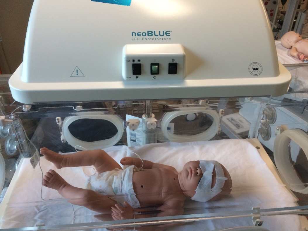
Figure 5: Proper setup for phototherapy treatment
Phototherapy is the primary treatment for hyperbilirubinemia. It works by converting unconjugated bilirubin in the skin to water-soluble isomers that can be excreted without liver conjugation.
Nursing Responsibilities for Phototherapy:
- Preparation and setup:
- Verify physician’s order and bilirubin levels
- Prepare equipment (phototherapy unit, eye protection, temperature probe)
- Position lights correctly (20-30 cm from infant for conventional; as per manufacturer’s guidelines for LED/fiberoptic)
- Measure irradiance (optimally 30+ μW/cm²/nm)
- Patient care during phototherapy:
- Place infant naked except for eye protection and diaper
- Ensure proper eye protection (cover eyes completely, check for pressure areas)
- Monitor vital signs q4h or as ordered
- Monitor temperature closely (risk of hypothermia or hyperthermia)
- Reposition infant every 2 hours for even light exposure
- Remove from phototherapy and remove eye patches during feeding
- Monitor skin for rashes, pressure areas, or burns
- Fluid management:
- Increase feeding frequency (phototherapy increases insensible water loss)
- Monitor intake and output (aim for urine output 1-3 mL/kg/hour)
- Weigh daily (assess for dehydration)
- Monitor for loose, greenish stools (common during phototherapy)
- Monitoring effectiveness:
- Check serum bilirubin levels as ordered (typically every 4-12 hours)
- Document response to treatment
- Continue phototherapy until bilirubin reaches safe level (typically 2-3 mg/dL below threshold for starting therapy)
- Obtain rebound bilirubin level 12-24 hours after discontinuation of phototherapy
- Parent education and support:
- Explain purpose and process of phototherapy
- Address parental concerns about separation from infant
- Teach parents about continued monitoring for jaundice after therapy
Types of Phototherapy
- Conventional phototherapy: Fluorescent or halogen lamps positioned above the infant
- Intensive phototherapy: Light sources above and below the infant or multiple light sources
- Fiberoptic blankets: Portable light source through fiberoptic pad (allows holding and breastfeeding during treatment)
- LED phototherapy: More efficient, generates less heat, longer lifespan
Exchange Transfusion
Exchange transfusion is used for severe hyperbilirubinemia when phototherapy is inadequate or bilirubin levels are approaching toxic thresholds. It involves removing the infant’s blood in small aliquots and replacing it with donor blood.
Nursing Responsibilities for Exchange Transfusion:
- Preparation:
- Verify blood product (type, crossmatch, expiration)
- Prepare equipment (umbilical catheters, syringes, monitoring devices)
- Obtain baseline vital signs and laboratory values
- Ensure emergency medications are available
- Place infant NPO 3-4 hours before procedure
- During procedure:
- Assist physician with procedure
- Monitor vital signs continuously (heart rate, respiratory rate, blood pressure, temperature, oxygen saturation)
- Watch for signs of complications (bradycardia, hypotension, hypocalcemia)
- Document volume of blood removed and replaced
- Maintain accurate record of laboratory specimens
- Post-procedure care:
- Continue phototherapy as ordered
- Monitor vital signs frequently (initially every 15-30 minutes)
- Check bilirubin levels 1-2 hours after procedure
- Monitor for hypoglycemia, hypocalcemia, and acid-base disturbances
- Observe for bleeding from catheter sites
- Resume feeding when stable
Potential Complications of Exchange Transfusion
- Hypocalcemia (due to citrate in donor blood)
- Thrombocytopenia
- Electrolyte imbalances
- Acid-base disturbances
- Cardiac arrhythmias
- Necrotizing enterocolitis
- Infection
- Thrombosis/embolism
- Volume overload or depletion
- Graft-versus-host disease
Supportive Care
Supportive care is essential for all infants with hyperbilirubinemia, particularly focusing on adequate nutrition, hydration, and monitoring.
Nutritional Support:
- Encourage frequent breastfeeding (8-12 times in 24 hours)
- Assist mothers with proper positioning and latch techniques
- Monitor for effective feeding (audible swallowing, adequate latch)
- Consider supplemental feeding if weight loss exceeds 10% or dehydration is present
- Avoid routine water or dextrose water supplementation (dilutes calories and may worsen jaundice)
- For breastfed infants with significant jaundice, continue breastfeeding (temporary interruption is rarely indicated)
Hydration Management:
- Ensure adequate hydration to facilitate bilirubin excretion
- Monitor weight daily (concern if loss >10% of birth weight)
- Track intake and output (aim for 6+ wet diapers per day after day 4)
- Assess for signs of dehydration (dry mucous membranes, sunken fontanelle, decreased skin turgor)
- For infants under phototherapy, increase fluid intake by 10-15% to compensate for increased insensible water loss
- IV fluids may be necessary if oral intake is inadequate or dehydration is present
Thermoregulation:
- Monitor body temperature frequently during phototherapy
- Maintain neutral thermal environment (risk of both hypothermia and hyperthermia during phototherapy)
- Adjust incubator temperature or distance of phototherapy lights as needed
- Monitor for signs of cold stress (increased oxygen consumption can worsen jaundice)
Pharmacological Management:
- Intravenous immunoglobulin (IVIG) for hemolytic disease (ABO or Rh incompatibility)
- Phenobarbital (rarely used; may increase bilirubin conjugation)
- Metalloporphyrins (experimental; inhibit heme oxygenase to reduce bilirubin production)
- Calcium supplementation during exchange transfusion to prevent hypocalcemia
Nursing Care Plan
| Nursing Diagnosis | Interventions | Expected Outcomes |
|---|---|---|
| Risk for Injury related to elevated bilirubin levels |
|
|
| Risk for Imbalanced Fluid Volume related to increased insensible water loss during phototherapy |
|
|
| Ineffective Thermoregulation related to phototherapy treatment |
|
|
| Risk for Impaired Skin Integrity related to exposure to phototherapy lights |
|
|
| Interrupted Breastfeeding related to infant’s condition and treatment |
|
|
| Deficient Knowledge (Parental) related to unfamiliarity with jaundice and its treatment |
|
|
Educational Topics for Parents
- Explanation of jaundice and its causes
- Treatment options and their rationale
- Importance of feeding for bilirubin excretion
- Signs and symptoms requiring medical attention
- Home phototherapy management (if applicable)
- Follow-up appointments and testing
- Long-term prognosis and monitoring
Complications: Kernicterus
Kernicterus is the most serious complication of severe hyperbilirubinemia. It refers to bilirubin-induced neurological damage resulting from the deposition of unconjugated bilirubin in brain tissue, particularly the basal ganglia and brainstem nuclei.
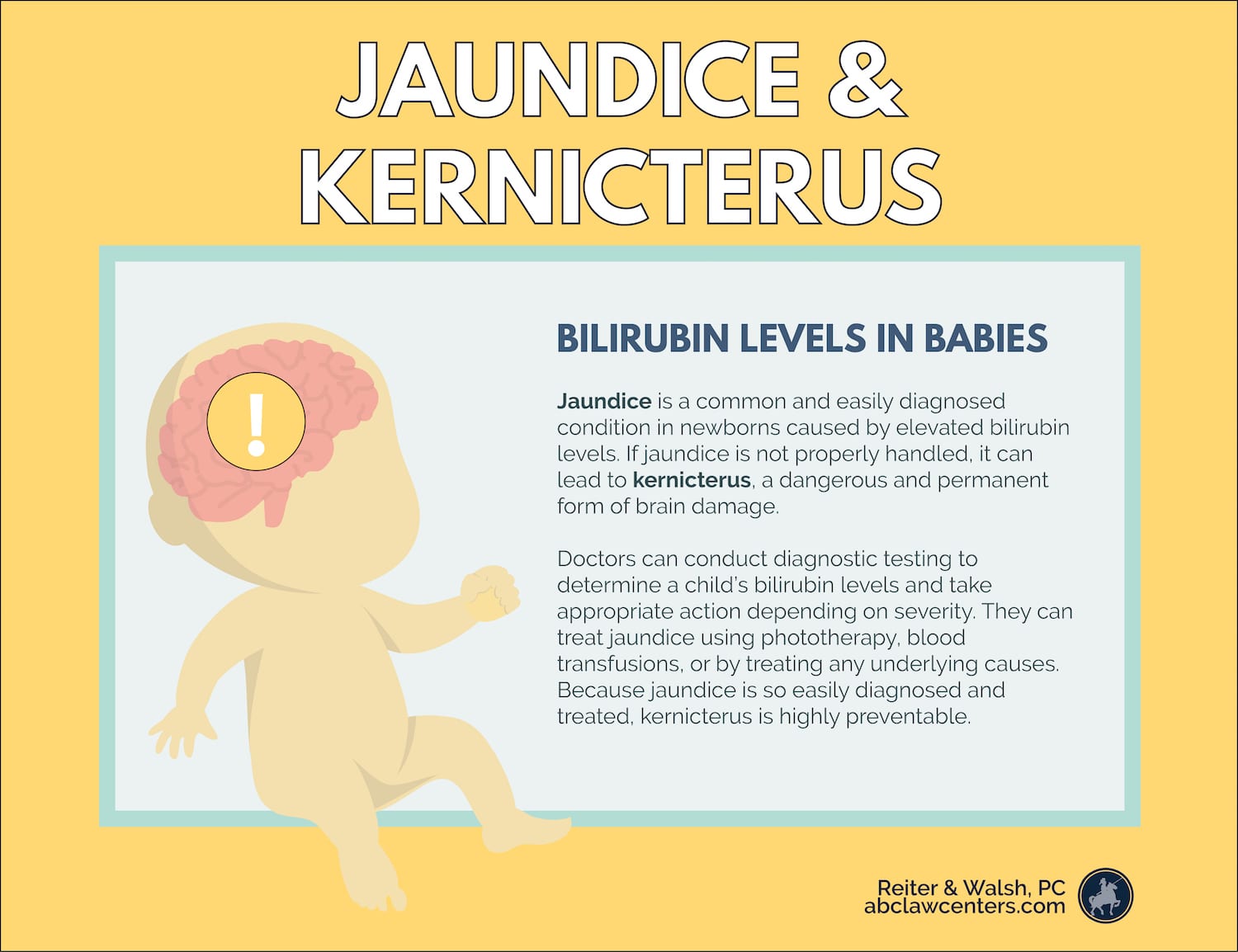
Figure 6: Progression from hyperbilirubinemia to kernicterus with neurological effects
Pathophysiology of Kernicterus
- Blood-brain barrier penetration: Unconjugated bilirubin, when not bound to albumin, can cross the blood-brain barrier.
- Neuronal damage: Bilirubin is toxic to neuronal cells, causing:
- Disruption of cellular membrane function
- Inhibition of mitochondrial enzymes
- Interference with neurotransmitter synthesis and release
- Inhibition of protein phosphorylation
- Disruption of DNA synthesis
- Preferential damage: Specifically affects the basal ganglia, hippocampus, cerebellum, and cranial nerve nuclei.
- Progression: Initially causes reversible brain injury (acute bilirubin encephalopathy), which can progress to permanent damage (kernicterus) if untreated.
Clinical Manifestations of Bilirubin Encephalopathy
| Phase | Clinical Signs | Nursing Implications |
|---|---|---|
| Phase 1 (Early) |
|
|
| Phase 2 (Intermediate) |
|
|
| Phase 3 (Advanced) |
|
|
| Chronic Kernicterus (>1 year) |
|
|
Prevention of Kernicterus: The Nurse’s Role
- Early identification of at-risk infants: Recognize risk factors and monitor bilirubin levels accordingly
- Systematic assessment: Monitor all infants for jaundice at least every 8-12 hours in the first 48 hours
- Prompt intervention: Initiate phototherapy according to guidelines based on bilirubin levels and risk factors
- Neurological monitoring: Perform frequent neurological assessments to detect early signs of bilirubin encephalopathy
- Parent education: Teach parents to recognize signs of jaundice and when to seek medical attention
- Appropriate follow-up: Ensure high-risk infants have timely follow-up appointments after discharge
Long-term Follow-up for Infants with Severe Hyperbilirubinemia
Infants who have experienced severe hyperbilirubinemia should be monitored for potential sequelae, including:
- Hearing assessment (before 3 months of age)
- Neurological evaluation (including motor development)
- Developmental screening
- Vision assessment
- Language development monitoring
Early intervention services should be initiated if developmental concerns are identified, as early intervention can significantly improve outcomes.
Review and Key Points
BILIRUBIN Mnemonic for Nursing Management
B – Blood tests to monitor bilirubin levels and assess for hemolysis
I – Identify risk factors and early signs of hyperbilirubinemia
L – Light therapy (phototherapy) implementation and monitoring
I – Intake and output monitoring to prevent dehydration
R – Regular assessment of neurological status for signs of encephalopathy
U – Understand and implement proper positioning during phototherapy
B – Breastfeeding support and nutritional management
I – Inform and educate parents about jaundice and its management
N – Note changes in condition and respond appropriately
Essential Knowledge Review
-
Physiology and Pathophysiology
- Bilirubin is primarily produced from the breakdown of hemoglobin
- Unconjugated bilirubin is lipid-soluble and potentially neurotoxic
- Conjugated bilirubin is water-soluble and can be excreted
- Neonates have physiological limitations in bilirubin metabolism
-
Types of Jaundice
- Physiological jaundice: Appears after 24 hours, peaks at 3-5 days, resolves by 7-10 days
- Pathological jaundice: Appears within 24 hours, rises rapidly, or persists beyond 2 weeks
- Breast feeding jaundice: Related to inadequate intake in first few days
- Breast milk jaundice: Later onset, related to factors in breast milk
-
Risk Factors
- Prematurity, ABO/Rh incompatibility, G6PD deficiency, significant bruising
- Inadequate feeding, weight loss >10%, dehydration
- Previous sibling with jaundice, certain ethnicities
-
Assessment
- Visual assessment using Kramer’s rule (cephalocaudal progression)
- Transcutaneous bilirubin (TcB) measurement for screening
- Total serum bilirubin (TSB) for definitive diagnosis and treatment decisions
- Risk assessment using hour-specific nomograms
-
Treatment Modalities
- Phototherapy: Primary treatment for most cases
- Exchange transfusion: For severe cases not responding to phototherapy
- IVIG: For immune-mediated hemolytic disease
- Supportive care: Adequate hydration and nutrition
-
Kernicterus
- Progression from acute bilirubin encephalopathy to permanent neurological damage
- Early signs: Lethargy, hypotonia, poor feeding, high-pitched cry
- Late signs: Hypertonia, retrocollis, opisthotonus, seizures
- Long-term sequelae: Choreoathetoid cerebral palsy, hearing loss, gaze abnormalities
-
Nursing Management
- Early identification of at-risk infants
- Promotion of adequate feeding and hydration
- Proper implementation of phototherapy
- Monitoring for signs of kernicterus
- Parent education and support
- Discharge planning and follow-up
Critical Thinking Challenges
Scenario 1: Preterm Infant with Jaundice
A 35-week gestational age infant is noted to have jaundice at 30 hours of life. The total serum bilirubin is 11 mg/dL. The infant is breastfeeding but has lost 9% of birth weight.
Questions to Consider:
- What risk factors does this infant have for developing severe hyperbilirubinemia?
- What assessments would you prioritize for this infant?
- What interventions would be appropriate at this time?
- What education would you provide to the parents?
Scenario 2: Term Infant Under Phototherapy
A 3-day-old term infant is receiving phototherapy for a total serum bilirubin of 18 mg/dL. The mother wants to continue exclusive breastfeeding but is concerned about the need to remove the baby from the lights for feeding.
Questions to Consider:
- How would you balance the need for phototherapy with the importance of breastfeeding?
- What signs of complications would you monitor for during phototherapy?
- What education would you provide to support the mother’s breastfeeding goals?
- What criteria would indicate that phototherapy can be discontinued?
References
- American Academy of Pediatrics Subcommittee on Hyperbilirubinemia. (2004). Management of hyperbilirubinemia in the newborn infant 35 or more weeks of gestation. Pediatrics, 114(1), 297-316.
- Bhutani, V. K., & Committee on Fetus and Newborn. (2011). Phototherapy to prevent severe neonatal hyperbilirubinemia in the newborn infant 35 or more weeks of gestation. Pediatrics, 128(4), e1046-e1052.
- Kaplan, M., Bromiker, R., & Hammerman, C. (2020). Hyperbilirubinemia, hemolysis, and increased bilirubin neurotoxicity. Seminars in Perinatology, 44(2), 151225.
- Maisels, M. J. (2015). Managing the jaundiced newborn: a persistent challenge. Canadian Medical Association Journal, 187(5), 335-343.
- Muchowski, K. E. (2014). Evaluation and treatment of neonatal hyperbilirubinemia. American Family Physician, 89(11), 873-878.
- Olusanya, B. O., Kaplan, M., & Hansen, T. W. R. (2018). Neonatal hyperbilirubinaemia: a global perspective. The Lancet Child & Adolescent Health, 2(8), 610-620.
- Rennie, J., Burman-Roy, S., & Murphy, M. S. (2010). Neonatal jaundice: summary of NICE guidance. BMJ, 340, c2409.
- Watchko, J. F., & Tiribelli, C. (2013). Bilirubin-induced neurologic damage—mechanisms and management approaches. New England Journal of Medicine, 369(21), 2021-2030.
- Wong, R. J., & Stevenson, D. K. (2015). Neonatal hemolysis and risk of bilirubin-induced neurologic dysfunction. Seminars in Fetal and Neonatal Medicine, 20(1), 26-30.