The Nurse’s Ultimate Guide to Patient Mobility and Immobility
Introduction: Why Mobility is a Pillar of Nursing Care
In nursing, movement is medicine, and immobility is the silent adversary. This simple truth underscores a core responsibility of every nurse. Promoting patient mobility and actively preventing the hazards of being bedridden are not just tasks on a checklist; they are fundamental interventions that influence everything from recovery speed to patient survival. For nursing students, mastering the principles of mobility is as critical as learning pharmacology or sterile technique.
This guide is designed in the “Osmosis-style”—a high-yield, clinically focused resource to help you master this competency. We will deconstruct the science of normal movement, explore the devastating systemic effects of immobility, and equip you with the evidence-based nursing interventions needed to manage both. Prepare to dive deep into a topic that is central to holistic, effective patient care.
The Science of Movement: Foundations for Practice
Before we can address impaired mobility, we must first understand the intricate coordination required for normal, purposeful movement. This foundation allows us to identify deviations and intervene effectively.
Elements of Normal Movement
Purposeful movement is not a single action but a symphony of multiple physiological components working in harmony. As defined by nursing fundamentals, these elements include alignment, joint mobility, and coordinated control.
Body Alignment & Posture
Body alignment refers to the relationship of one body part to another along a vertical or horizontal line. Correct alignment minimizes strain on joints, tendons, ligaments, and muscles. Good posture is the practical application of proper alignment, whether standing, sitting, or lying down, which maintains muscle tone and contributes significantly to balance. As described in clinical skills textbooks, when the body is well-aligned, the strain on musculoskeletal structures is minimized, reducing fatigue and the risk of injury.
Visual Aid: Posture Alignment
Correct Posture
Spine’s natural curves maintained. Center of gravity is stable.
Incorrect Posture
Exaggerated curves (slouching) strain muscles and ligaments.
Joint Mobility (Range of Motion – ROM)
Joint mobility is the maximum range of movement possible for a specific joint. This range is determined by the joint’s structure, ligament elasticity, and surrounding muscle condition. Key types of joint movements include:
- Flexion & Extension: Bending and straightening a joint (e.g., elbow, knee).
- Abduction & Adduction: Moving a limb away from or toward the body’s midline.
- Rotation: Turning a joint around its axis (e.g., neck, hip).
Maintaining joint mobility is crucial, as immobility can lead to stiffness and contractures in as little as three days.
Balance & Coordinated Movement
This complex process is governed by the nervous system. The cerebellum coordinates all voluntary movements, while the cerebral cortex initiates them. Balance is achieved when the body’s center of gravity is maintained over a stable base of support. A wide base of support (feet apart) and a low center of gravity (bending the knees) increase stability, a core principle in both patient and nurse safety.
Principles of Body Mechanics: Protecting Yourself and Your Patient
Musculoskeletal injuries (MSIs) are a leading occupational hazard in nursing. While modern healthcare emphasizes the use of Safe Patient Handling and Mobility (SPHM) technology like mechanical lifts, understanding proper body mechanics remains essential for situations where manual handling is unavoidable. The American Nurses Association (ANA) advocates for eliminating manual patient handling, but these principles reduce risk when it must occur.
Mnemonic for Body Mechanics: B.A.C.K.
Use this simple acronym to guide your movements:
- B – Back Straight: Keep your back straight; bend at your knees and hips.
- A – Avoid Twisting: Pivot your entire body by moving your feet.
- C – Close to Body: Hold the patient or object close to your center of gravity.
- K – Keep Smooth: Move in a smooth, coordinated manner, not with jerky motions.
Key principles include:
- Assess First: Evaluate the patient’s ability to assist, the environment (is it cluttered?), and whether you need help or equipment.
- Wide Base of Support: Place your feet shoulder-width apart to maximize stability.
- Work at Waist Level: Raise the bed to a comfortable working height to avoid stooping.
- Bend at the Knees: Use the strong muscles of your legs and glutes to do the lifting, not your back.
- Push, Don’t Pull: It generally requires less energy to push an object than to pull it.
When Movement is Compromised
Impaired physical mobility is rarely caused by a single issue. It is often a multifactorial problem stemming from a combination of physiological, psychological, and environmental conditions.
Factors Affecting Body Alignment and Activity
Understanding the root causes of immobility is the first step in creating an effective care plan. These factors can be broadly categorized.
Flowchart: Contributors to Impaired Mobility
- Physiological Factors
(Growth/Development, Physical Health like fractures, stroke, pain, fatigue, poor nutrition) - Psychological Factors
(Depression, Fear of Falling, Cognitive Impairment, Lack of Motivation) - External/Environmental Factors
(Prescribed Bed Rest, Lack of Assistive Devices, Unsafe Environment) - Result: Impaired Physical Mobility
As noted in nursing literature, even a patient’s mental state can profoundly impact their willingness to move. For many chronically ill patients, factors like pain, weakness, and fatigue create significant barriers to activity. A patient with depression may lack the motivation, while an elderly patient who has fallen once may develop a debilitating fear of falling again, leading to a cycle of decreased activity and further deconditioning.
Alterations in Mobility: The Nursing Diagnosis
In the nursing process, we frame these issues using a formal diagnosis. The most common is Impaired Physical Mobility.
According to NANDA-International (NANDA-I), this diagnosis is defined as a “Limitation in independent, purposeful physical movement of the body or of one or more extremities.”
To make this diagnosis clinically useful, we write it as a three-part PES statement (Problem, Etiology, Signs/Symptoms):
Problem: Impaired Physical Mobility
Etiology (Related to): decrease in muscle strength
Signs/Symptoms (As Evidenced By): slow movement and alteration in gait.
Problem: Impaired Physical Mobility
Etiology (Related to): pain and stiffness secondary to arthritis
Signs/Symptoms (As Evidenced By): reluctance to attempt movement and limited range of motion.
Crafting a precise PES statement is crucial because it directly guides your nursing interventions. If the cause is pain, the intervention is pain management. If it’s muscle weakness, the intervention is physical therapy and strengthening exercises.
The Cascade of Immobility: A System-by-System Breakdown
Prolonged immobility triggers a dangerous cascade of negative effects throughout the body. What starts as a simple restriction of movement can quickly evolve into a multi-system failure. Understanding this process is key to preventing it. As one nursing resource aptly puts it, immobility is “enemy #1” in patient care.
Integumentary System
- Effect: Pressure Ulcers (Pressure Injuries)
- Pathophysiology: Constant pressure on skin over bony prominences (e.g., sacrum, heels, hips) compresses capillaries, obstructing blood flow. This leads to tissue ischemia, and if unrelieved, necrosis and breakdown.
- Assessment: Inspect skin at least daily, especially over bony areas. Check for non-blanchable erythema (redness that doesn’t turn white when pressed), which is a Stage 1 pressure injury.
Musculoskeletal System
- Effect: Disuse Atrophy, Joint Contractures, Disuse Osteoporosis
- Pathophysiology: Without mechanical stress, muscles break down protein faster than they build it (atrophy). Unused joints develop shortening of connective tissues, leading to permanent fixation (contractures). Bones lose calcium due to lack of weight-bearing stress, becoming brittle and prone to fracture.
- Assessment: Assess muscle mass and strength (e.g., “push against my hands”). Measure passive and active ROM, noting any stiffness or pain.
Cardiovascular System
- Effect: Orthostatic Hypotension, Increased Cardiac Workload, Deep Vein Thrombosis (DVT)
- Pathophysiology: Immobility leads to a decrease in circulating blood volume and autonomic deconditioning, causing a sharp drop in blood pressure upon standing (orthostatic hypotension). The heart works harder in a supine position. Venous stasis (pooling of blood in the legs), combined with potential hypercoagulability and vessel wall injury (Virchow’s Triad), creates a high risk for DVT.
- Assessment: Measure BP lying, sitting, and standing. Assess for dizziness or syncope. Check for signs of DVT: unilateral leg swelling, pain, warmth, and redness.
Respiratory System
- Effect: Atelectasis & Hypostatic Pneumonia
- Pathophysiology: In a supine position, chest expansion is restricted, and secretions pool in the dependent areas of the lungs. This collapse of alveoli (atelectasis) creates a perfect breeding ground for bacteria, leading to hypostatic pneumonia.
- Assessment: Auscultate breath sounds for crackles or diminished sounds. Monitor respiratory rate, depth, and SpO2. Assess the effectiveness of the patient’s cough.
Gastrointestinal (GI) & Genitourinary (GU) Systems
- Effect: Constipation, Urinary Stasis, Renal Calculi (Kidney Stones)
- Pathophysiology: Decreased physical activity slows peristalsis, leading to constipation. Lying supine makes it difficult to empty the bladder completely (urinary stasis), increasing the risk for UTIs and the formation of renal calculi from concentrated urine.
- Assessment: Monitor frequency and consistency of bowel movements. Assess for urinary retention, potentially with a bladder scanner. Note urine color and clarity.
Metabolic & Psychological Systems
- Effect: Negative Nitrogen Balance, Insulin Resistance, Depression, Confusion
- Pathophysiology: Muscle breakdown releases more nitrogen than the body ingests through protein, leading to a negative nitrogen balance and impaired wound healing. Immobility can also decrease the body’s sensitivity to insulin. Sensory deprivation, loss of independence, and boredom contribute to depression, anxiety, and delirium.
- Assessment: Monitor nutritional intake and wound healing. Check blood glucose levels. Perform regular mental status exams and screen for depression.
The Nursing Process in Action: Promoting Mobility and Preventing Harm
Armed with knowledge of why mobility matters, the nurse’s role shifts to proactive intervention. This involves a continuous cycle of assessment, planning, intervention, and evaluation.
Assessment: The First Step to Safe Mobility
A thorough mobility assessment is the foundation of a safe and effective care plan. It goes beyond simply asking “Can you walk?”.
- Mobility Status: Systematically evaluate the patient’s ability in three key areas:
- Bed Mobility: Can the patient turn side-to-side, or move from lying to sitting?
- Transfers: How much assistance is needed to move from bed to chair? Can they stand?
- Ambulation: If they can walk, assess their gait, balance, endurance, and need for assistive devices.
- Range of Motion (ROM): Assess major joints (shoulders, elbows, hips, knees) for both active (patient moves) and passive (nurse moves) ROM. Note any limitations, pain, or crepitus.
- Muscle Strength: Test strength and equality in upper and lower extremities.
- Fall Risk: Use a validated tool, such as the Morse Fall Scale or Hendrich II Fall Risk Model, to quantify risk and guide precautions.
- Complication Assessment: Actively look for the signs and symptoms of immobility complications detailed in the previous section (e.g., skin inspection, lung auscultation, checking for DVT).
Interventions to Maintain and Restore Mobility
Exercise as a Therapeutic Tool
Exercise for hospitalized patients is not about athletic training; it’s about preventing deconditioning. The main types include:
| Exercise Type | Description | Benefits & Clinical Use |
|---|---|---|
| Isotonic | Active movement with muscle shortening (e.g., walking, performing ADLs, swimming). | Improves circulation, muscle tone, and joint mobility. The goal for most patients is to progress to this type of activity. |
| Isometric | Muscle contraction without joint movement (e.g., tightening quadriceps or gluteal muscles and holding for 10 seconds). | Maintains muscle strength and tone. Ideal for patients on strict bed rest or those in casts. |
| Range-of-Motion (ROM) | Moving a joint through its full range. Can be Active (patient performs) or Passive (nurse performs). | Prevents joint contractures and stiffness. Passive ROM does not build muscle but maintains flexibility. Simple bed exercises like ankle circles and heel slides are excellent examples. |
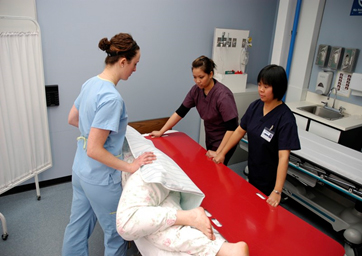
Safe Patient Handling, Positioning, and Transfers
How we move and position patients is a critical intervention. The goal is to maintain alignment, promote comfort, and prevent complications.
Positioning: A frequent turning and repositioning schedule (typically every 2 hours) is the cornerstone of pressure injury prevention. Key therapeutic positions include:
Common Patient Positions
Fowler’s Position (45-60°)
Use: Promotes lung expansion, decreases intracranial pressure. Ideal for patients with respiratory distress or NGT.
Supine Position
Use: Post-operative recovery, general examination. Requires careful padding of bony prominences.
Prone Position
Use: Promotes drainage of oral secretions, prevents hip flexion contractures. Used in ARDS management.
Lateral (Side-Lying)
Use: Relieves pressure on the sacrum. A 30-degree lateral incline is recommended for pressure injury prevention.
For detailed guidelines on each position, refer to a comprehensive guide like the one from Nurseslabs.
Transfers & Ambulation: The goal is to move the patient safely while encouraging them to participate as much as possible. This maintains their strength and dignity. Assistive devices are crucial for safety.
- Gait Belt: Provides a secure point of contact for the nurse to support the patient’s center of gravity during transfers or ambulation.
- Walker/Cane: Increases the patient’s base of support, improving stability.
- Mechanical Lifts: Essential for non-weight-bearing or unpredictable patients. Using these devices is a key component of SPHM programs and protects both the patient and the nurse from injury.
- Slider Boards: Reduce friction during lateral transfers (e.g., from a stretcher to a bed), minimizing shear forces on the patient’s skin.
Interventions to Prevent Complications of Immobility
Prevention is always the best approach. Your interventions should be targeted at the specific risks of immobility.
- Pressure Ulcer Prevention: Implement a consistent 2-hour turning schedule. Use pressure-redistributing surfaces (specialty mattresses). Keep skin clean and dry, and apply moisture barriers as needed. Ensure adequate protein and hydration.
- DVT Prevention: Encourage active leg exercises like ankle pumps. Apply sequential compression devices (SCDs) or anti-embolism stockings as ordered. Administer prophylactic anticoagulants (e.g., heparin, enoxaparin) precisely as prescribed.
- Contracture Prevention: Perform passive or active ROM exercises multiple times a day. Use positioning aids like hand rolls and foot boots to maintain functional alignment.
- Pneumonia Prevention: Encourage the patient to cough, take deep breaths, and use an incentive spirometer every 1-2 hours while awake. Early mobilization—even just sitting in a chair—is highly effective.
Putting It All Together: Clinical Tools and Takeaways
Mastering this topic requires integrating knowledge into daily practice. These tools are designed for quick recall and application at the bedside.
Quick-Reference Memory Aids
Mnemonic for Immobility Complications: BEDSORES
Use this acronym to quickly recall the major systemic risks:
- B – Blood clots (DVT/PE)
- E – Emotional distress (Depression/Confusion)
- D – Decreased lung expansion (Atelectasis/Pneumonia)
- S – Skin breakdown (Pressure Ulcers)
- O – Osteoporosis (Bone demineralization)
- R – Renal issues (Stasis/Calculi)
- E – Energy loss (Muscle Atrophy)
- S – Stool impaction (Constipation)
- Assess Mobility & Fall Risk on Admission: Establish a baseline immediately.
- Turn and Reposition Every 2 Hours: Non-negotiable for bed-bound patients.
- Mobilize Early and Often: Get the patient out of bed to a chair or ambulating as soon as safely possible.
- Encourage Deep Breathing & Coughing: Actively prevent pneumonia.
- Ensure Adequate Hydration & Nutrition: The body cannot heal or maintain tissue integrity without fuel.
Sample Nursing Note: Documenting Mobility
Clear, concise documentation is essential for communication and continuity of care. It should paint a picture of the patient’s functional status and the interventions performed.
Narrative Note Example:
“Patient assessed for mobility. Able to reposition self in bed with use of overhead trapeze. Requires assistance of 1 for transfer from bed to chair using a walker; gait is slow and steady for 20 feet before reporting fatigue. Denies dizziness or pain during ambulation. Lungs clear to auscultation bilaterally post-use of incentive spirometer. Skin intact over sacrum and heels, no redness noted. Sequential compression devices in place and functioning while in bed. Actively participating in ankle pump exercises. Fall precautions remain in place. Plan: Continue to encourage ambulation in halls TID with assistance.”
Conclusion: The Proactive Nurse
Mobility should be considered a vital sign. It reflects and affects the health of every body system. A proactive nurse does not wait for complications to arise; they anticipate them. By mastering the assessment of mobility, understanding the cascade of immobility, and consistently applying preventative interventions, you become a true champion of movement. This proactive approach can prevent a cascade of complications, shorten hospital stays, reduce healthcare costs, and, most importantly, dramatically improve your patients’ outcomes and quality of life.