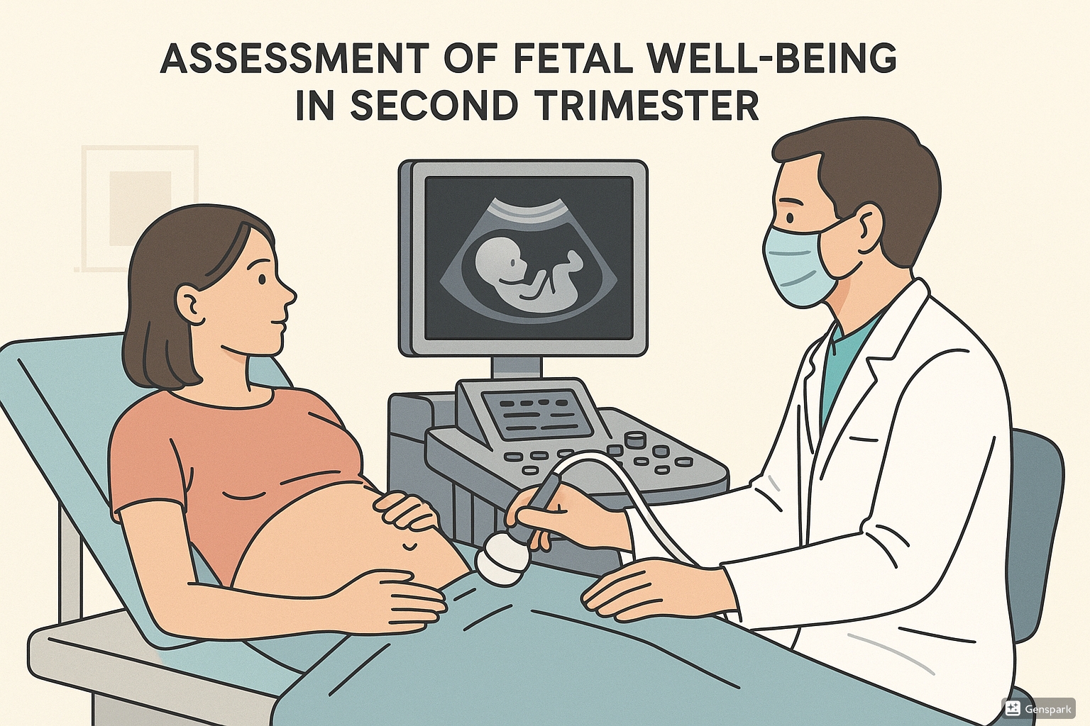
Assessment of Fetal Well-being in Second Trimester
A Comprehensive Guide for Nursing Students

Table of Contents
1. Introduction to Fetal Well-being Assessment
Fetal well-being assessment is a critical component of prenatal care, especially during the second trimester (14-28 weeks). These assessments aim to identify fetuses at risk for intrauterine distress, growth restriction, or death, enabling timely interventions to improve outcomes. The assessment techniques range from simple maternal perception of movements to sophisticated technological and biochemical evaluations.
In the second trimester, several physiological milestones make assessment particularly valuable:
- Fetal movement becomes perceptible to the mother (quickening)
- Fetal heart tones can be reliably detected
- Anatomical structures are well-formed and assessable via ultrasound
- Maternal-fetal circulation is well-established
2. Daily Fetal Movement Count (DFMC)
Daily Fetal Movement Count (DFMC) is one of the simplest, most cost-effective methods for assessing fetal well-being. It relies on maternal perception of fetal movements as an indicator of fetal health.
Technique
The DFMC technique involves:
- Recording fetal movements perceived by the mother for one hour after meals (breakfast, lunch, and dinner)
- Counting distinct movements (kicks, rolls, flutters)
- Maintaining a daily chart of these movements
Interpretation
| Count | Interpretation | Action |
|---|---|---|
| ≥3 movements per hour | Satisfactory/Reassuring | Continue routine monitoring |
| ≥10 movements in 12 hours | Normal | Continue routine monitoring |
| <3 movements per hour | Concerning/Non-reassuring | Further assessment needed |
| Absence of movements | Alarming | Immediate medical evaluation |
3. Biophysical Profile (BPP)
The Biophysical Profile (BPP) is a comprehensive assessment of fetal well-being that evaluates five parameters, four by ultrasound and one by non-stress test. Though typically used in the third trimester, it may be initiated in the late second trimester in high-risk pregnancies.
Components of BPP
| Parameter | Normal Finding (2 points) | Abnormal Finding (0 points) |
|---|---|---|
| Fetal Breathing Movements | ≥1 episode of rhythmic breathing movements lasting ≥30 seconds in 30 minutes | Absent or brief breathing movements |
| Gross Body Movements | ≥3 discrete body/limb movements in 30 minutes | <3 movements in 30 minutes |
| Fetal Tone | ≥1 episode of extension with return to flexion of fetal limbs or trunk | Slow extension with partial return or movement absence |
| Amniotic Fluid Volume | ≥1 pocket of amniotic fluid measuring ≥2cm in perpendicular planes | No or small pockets of amniotic fluid |
| Non-stress Test (NST) | ≥2 accelerations of ≥15 beats/min lasting ≥15 seconds in 20 minutes | 0 or 1 acceleration in 20 minutes |
BPP Score Interpretation
| Score | Interpretation | Management |
|---|---|---|
| 8-10 | Normal | Routine follow-up |
| 6 | Equivocal | Repeat test within 24 hours |
| 4 | Suspicious | Further evaluation needed, consideration for delivery |
| 0-2 | Abnormal | Immediate delivery if fetus viable |
4. Non-Stress Test (NST)
The Non-Stress Test (NST) evaluates fetal heart rate (FHR) response to fetal movement, reflecting the integrity of the fetal autonomic nervous system and cardiac function. It can be performed as early as 24-26 weeks gestation in the second trimester.
Procedure
- Position patient in semi-Fowler’s or left lateral position
- Apply external electronic fetal monitor with tocodynamometer
- Observe and record FHR for 20-30 minutes
- Ask mother to press event marker button when fetal movement is felt
- Document contractions, baseline FHR, variability, accelerations, and decelerations
Interpretation
| Result | Criteria | Interpretation |
|---|---|---|
| Reactive (Normal) | ≥2 FHR accelerations of ≥15 beats/min lasting ≥15 seconds in 20 minutes | Reassuring fetal status |
| Non-reactive | Insufficient accelerations as defined above | May indicate fetal compromise or sleep cycle |
| Unsatisfactory | Poor quality tracing, insufficient monitoring time | Repeat test |
5. Cardiotocography (CTG)
Cardiotocography (CTG) is a technical method of recording the fetal heartbeat and uterine contractions simultaneously. Though more commonly used in the third trimester, it can be valuable in the late second trimester (after 24 weeks) when concerning symptoms arise.
Technical Aspects
CTG uses two transducers placed on the mother’s abdomen:
- Ultrasound transducer: Records fetal heart rate using Doppler ultrasound principles
- Pressure transducer: Records uterine contractions (tocodynamometer)
Parameters Assessed
| Parameter | Normal Range | Abnormal Findings |
|---|---|---|
| Baseline FHR | 110-160 bpm | Bradycardia (<110 bpm) or Tachycardia (>160 bpm) |
| Variability | 5-25 bpm | Reduced (<5 bpm) or Absent (0 bpm) |
| Accelerations | Present with fetal movement | Absent |
| Decelerations | Absent | Early, Late, or Variable decelerations |
| Contractions | 0-5 in 10 minutes | >5 in 10 minutes (tachysystole) |
6. Ultrasound (USG) Assessment
Ultrasound is a cornerstone in the assessment of fetal well-being during the second trimester. It provides detailed information about fetal anatomy, growth, and development without ionizing radiation.
Key Parameters Assessed
- Fetal growth parameters:
- Biparietal diameter (BPD)
- Head circumference (HC)
- Abdominal circumference (AC)
- Femur length (FL)
- Estimated fetal weight (EFW)
- Amniotic fluid assessment:
- Single deepest pocket (SDP)
- Amniotic fluid index (AFI)
- Placental assessment:
- Location and morphology
- Placental grade
- Doppler ultrasound:
- Umbilical artery blood flow
- Middle cerebral artery blood flow
- Ductus venosus flow (in high-risk cases)
Amniotic Fluid Assessment Norms
| Parameter | Normal Range | Clinical Significance |
|---|---|---|
| Amniotic Fluid Index (AFI) | 8-24 cm | <8 cm: Oligohydramnios >24 cm: Polyhydramnios |
| Single Deepest Pocket (SDP) | 2-8 cm | <2 cm: Oligohydramnios >8 cm: Polyhydramnios |
7. Vibroacoustic Stimulation
Vibroacoustic stimulation (VAS) is a simple, non-invasive technique to assess fetal response to external stimuli, particularly useful when combined with NST to improve test efficiency.
Procedure
- Place an electronic artificial larynx (vibroacoustic stimulator) on the maternal abdomen over the fetal head
- Activate the device to produce a sound stimulus (usually 80-90 dB) for 1-2 seconds
- Observe the fetal heart rate response
- The stimulation may be repeated up to 3 times if no response is obtained
Interpretation
| Response | Interpretation |
|---|---|
| FHR acceleration within 10 seconds | Reassuring (normal response) |
| No FHR acceleration after 3 stimulations | Non-reassuring (may indicate fetal compromise) |
Clinical Applications
- Shortening the duration of non-stress tests
- Stimulating fetal movement during biophysical profile assessment
- Differentiating between fetal sleep and compromise in non-reactive NSTs
8. Biochemical Tests
Biochemical markers provide valuable insights into fetal well-being and development during the second trimester. These tests can help identify potential chromosomal abnormalities, neural tube defects, and other conditions that may affect fetal health.
Second Trimester Biochemical Markers
| Marker | Clinical Significance | Normal Range (MoM*) |
|---|---|---|
| Alpha-Fetoprotein (AFP) | Elevated: Neural tube defects, abdominal wall defects Decreased: Down syndrome |
0.5-2.5 |
| Unconjugated Estriol (uE3) | Decreased: Down syndrome, trisomy 18, IUGR | 0.5-2.0 |
| Human Chorionic Gonadotropin (hCG) | Elevated: Down syndrome Decreased: Trisomy 18 |
0.5-2.0 |
| Inhibin A | Elevated: Down syndrome | 0.5-2.0 |
*MoM = Multiple of the Median
Screening Test Combinations
- Triple Test: AFP + uE3 + hCG
- Detection rate for Down syndrome: 60-70%
- Quadruple (Quad) Test: AFP + uE3 + hCG + Inhibin A
- Detection rate for Down syndrome: 75-80%
- Optimal timing: 15-22 weeks (ideally 16-18 weeks)
Emerging Biochemical Markers
- Cell-free fetal DNA in maternal blood: Non-invasive prenatal testing with high sensitivity and specificity for common aneuploidies
- Placental Growth Factor (PlGF): Decreased levels may indicate risk for preeclampsia or fetal growth restriction
- Pregnancy-Associated Plasma Protein-A (PAPP-A): Low second-trimester levels may indicate risk for adverse pregnancy outcomes
9. Best Practices & Updates
Recent advancements and best practices in fetal well-being assessment have improved outcomes and patient experience. Here are three key updates that nursing professionals should be aware of:
1. Integration of Multiple Assessment Modalities
Current best practice emphasizes using multiple complementary assessment techniques rather than relying on a single method. This integrated approach provides more accurate evaluation of fetal status and reduces false positive/negative results.
Example: Combining maternal DFMC monitoring with periodic biophysical assessments has shown greater sensitivity in detecting early signs of fetal compromise compared to either technique alone.
2. Personalized Assessment Schedules
Recent guidelines recommend tailoring the frequency and type of fetal assessments based on individual risk profiles rather than applying a one-size-fits-all approach.
High-risk pregnancies (e.g., maternal hypertension, diabetes, previous stillbirth) may require BPP assessments beginning as early as 26-28 weeks, while low-risk pregnancies may focus primarily on maternal perception of movement with clinical validation.
3. Technological Advancements in Monitoring
New developments in fetal monitoring technology have enhanced our ability to assess fetal well-being with greater precision and convenience.
Remote monitoring solutions now allow pregnant women to record fetal movements and heart rates at home using smartphone-connected devices, with data transmitted directly to healthcare providers for review. This has shown particular benefit during the COVID-19 pandemic by reducing in-person visits while maintaining surveillance.
10. Clinical Applications
As nursing professionals, understanding when and how to apply different fetal well-being assessment techniques is crucial for optimal patient care.
Indications for Fetal Well-being Assessment in the Second Trimester
| Maternal Factors | Fetal Factors | Pregnancy Factors |
|---|---|---|
|
|
|
Nursing Role in Fetal Well-being Assessment
- Patient Education: Teaching pregnant women how to monitor fetal movements and when to seek medical attention
- Procedure Assistance: Preparing equipment, positioning patients, and assisting during ultrasound, NST, and BPP
- Monitoring: Recognizing normal and abnormal patterns during electronic fetal monitoring
- Documentation: Accurately recording all findings and communicating results to the healthcare team
- Support: Providing emotional support to patients, particularly when abnormal findings generate anxiety
Decision-Making Algorithm for Decreased Fetal Movement
- Perform focused history and assessment
- Initiate electronic fetal monitoring (NST)
- If non-reactive NST, proceed to BPP
- Normal BPP (≥8/10): Continue routine prenatal care with increased surveillance
- Abnormal BPP (≤6/10): Consider further testing and possible delivery planning based on gestational age and clinical context
