
Cysts and Tumors
Comprehensive Nursing Guide
Evidence-Based Clinical Practice
Table of Contents
Introduction
Cysts and tumors represent fundamental concepts in pathology that every nursing professional must thoroughly understand. These abnormal tissue growths affect millions of patients worldwide and require skilled nursing assessment, intervention, and management. Understanding the distinctions between these conditions is crucial for providing safe, effective, and compassionate patient care.
As healthcare providers on the front lines of patient care, nurses play a pivotal role in early detection, patient education, and comprehensive management of both benign and malignant growths. This comprehensive guide provides evidence-based information to enhance clinical decision-making and improve patient outcomes.
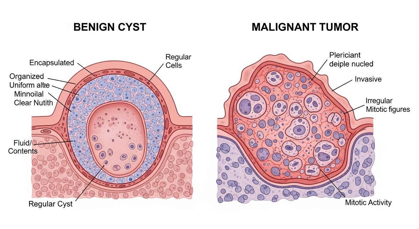
Figure 1: Anatomical comparison between benign cysts and malignant tumors showing cellular and structural differences
Definitions
Cysts
A cyst is a closed sac-like structure containing fluid, semi-solid material, or gas. Cysts are typically benign and characterized by:
- Well-defined boundaries
- Epithelial lining
- Non-neoplastic nature
- Slow growth pattern
- Usually painless unless infected or ruptured
Tumors
A tumor (neoplasm) is an abnormal mass of tissue resulting from uncontrolled cell proliferation. Tumors are characterized by:
- Autonomous growth
- Can be benign or malignant
- Variable growth rates
- Potential for metastasis (malignant)
- May cause local or systemic effects
Key Differences Summary
| Characteristic | Cysts | Tumors |
|---|---|---|
| Structure | Hollow, fluid-filled | Solid tissue mass |
| Growth Pattern | Expansion by accumulation | Cell proliferation |
| Malignant Potential | Rarely malignant | Can be benign or malignant |
| Symptoms | Often asymptomatic | Variable, may cause systemic effects |
Types and Classification
Cyst Classification
Congenital Cysts
- • Branchial cleft cysts
- • Thyroglossal duct cysts
- • Dermoid cysts
- • Polycystic kidney disease
Acquired Cysts
- • Sebaceous cysts
- • Ganglion cysts
- • Ovarian cysts
- • Baker’s cysts
Retention Cysts
- • Mucus retention cysts
- • Nabothian cysts
- • Chalazion
- • Ranula
Tumor Classification
Benign Tumors
Characteristics:
- Well-differentiated cells
- Slow growth
- Encapsulated
- No metastasis
- Minimal tissue invasion
Examples:
- • Lipomas
- • Fibromas
- • Adenomas
- • Leiomyomas
Malignant Tumors
Characteristics:
- Poorly differentiated cells
- Rapid growth
- Not encapsulated
- Metastatic potential
- Invasive growth pattern
Categories:
- • Carcinomas (epithelial)
- • Sarcomas (connective tissue)
- • Lymphomas (lymphatic)
- • Leukemias (blood)
Memory Aid: “SLIM vs RAPID”
Benign – SLIM:
- Slow growth
- Localized (encapsulated)
- Inert (no metastasis)
- Mild symptoms
Malignant – RAPID:
- Rapid growth
- Aggressive invasion
- Poor differentiation
- Invasive metastasis
- Dangerous systemic effects
Etiological Factors
Cyst Development Factors
Developmental Factors
- • Embryonic developmental anomalies
- • Incomplete organ development
- • Vestigial structure persistence
Obstruction-Related
- • Ductal blockage
- • Secretion accumulation
- • Inflammatory scarring
Hormonal Influences
- • Ovarian hormone fluctuations
- • Sebaceous gland stimulation
- • Growth factor imbalances
Infectious Causes
- • Parasitic infections (echinococcus)
- • Bacterial abscesses
- • Viral-induced formations
Tumor Development Factors
Genetic Factors
- • Oncogene activation
- • Tumor suppressor gene mutations
- • DNA repair gene defects
- • Inherited cancer syndromes
Environmental Carcinogens
- • Chemical exposure (benzene, asbestos)
- • Radiation (UV, ionizing)
- • Tobacco and alcohol
- • Occupational hazards
Infectious Agents
- • Human papillomavirus (HPV)
- • Hepatitis B and C viruses
- • Epstein-Barr virus
- • Helicobacter pylori
Lifestyle Factors
- • Diet and nutrition
- • Physical inactivity
- • Obesity
- • Chronic inflammation
Pathophysiology
Cyst Formation Mechanisms
Initial Stimulus
Obstruction, inflammation, or developmental anomaly
Secretion Accumulation
Fluid or material builds up behind obstruction
Cavity Formation
Expansion creates epithelial-lined sac
Mature Cyst
Stable, encapsulated fluid-filled structure
Cellular Characteristics
- • Epithelial lining maintains secretory function
- • Normal cellular differentiation
- • Minimal mitotic activity
- • Preserved cell-to-cell adhesion
Growth Pattern
- • Expansion by accumulation, not proliferation
- • Growth rate depends on secretion rate
- • Pressure-limited expansion
- • Self-limiting growth potential
Tumor Development Mechanisms
Benign Tumor Pathophysiology
- Cell Cycle Control: Maintained growth regulation
- Differentiation: Cells resemble tissue of origin
- Apoptosis: Normal programmed cell death
- Angiogenesis: Limited blood vessel formation
- Growth Factors: Respond to growth inhibitory signals
- Encapsulation: Surrounded by fibrous capsule
Malignant Tumor Pathophysiology
- Oncogene Activation: Uncontrolled growth signals
- Tumor Suppressor Loss: Loss of growth brakes
- Anaplasia: Loss of cellular differentiation
- Immortalization: Unlimited replicative potential
- Angiogenesis: Enhanced blood supply formation
- Invasion: Breakdown of basement membrane
- Metastasis: Spread to distant sites
Hallmarks of Malignant Transformation
Self-Sufficiency
Growth signal independence
Insensitivity
Ignore growth inhibitory signals
Apoptosis Evasion
Resistance to programmed death
Limitless Replication
Unlimited proliferative potential
Sustained Angiogenesis
Promote blood vessel formation
Tissue Invasion
Metastasis capability
Signs and Symptoms
Cyst Manifestations
General Characteristics
- • Often asymptomatic
- • Smooth, well-defined borders
- • Mobile unless adherent
- • Fluctuant or soft consistency
- • Size varies from millimeters to centimeters
Symptomatic Presentations
- • Cosmetic concerns
- • Pressure symptoms on adjacent structures
- • Pain if infected or ruptured
- • Functional impairment (location-dependent)
- • Intermittent swelling
Complications
- • Secondary infection
- • Rupture with inflammatory reaction
- • Hemorrhage into cyst cavity
- • Torsion (ovarian cysts)
- • Malignant transformation (rare)
Tumor Manifestations
Benign Tumor Signs
- • Slow, progressive growth
- • Well-circumscribed borders
- • Mobile mass
- • Painless unless pressure-related
- • No constitutional symptoms
- • Function preservation
Malignant Tumor Signs
- • Rapid growth pattern
- • Irregular, fixed borders
- • Hard, immobile mass
- • Ulceration or skin changes
- • Lymph node enlargement
- • Local tissue destruction
Systemic Symptoms
- • Unexplained weight loss
- • Fatigue and weakness
- • Night sweats
- • Loss of appetite
- • Fever of unknown origin
- • Paraneoplastic syndromes
Red Flag Symptoms Requiring Immediate Attention
- Rapid size increase
- Hard, fixed mass
- Irregular borders
- Skin changes or ulceration
- Lymphadenopathy
- Constitutional symptoms
- Functional impairment
- Pain not related to trauma
Assessment
Comprehensive Nursing Assessment
Subjective Assessment
History of Present Illness
- • Onset and duration
- • Growth pattern and rate
- • Associated symptoms
- • Pain characteristics (PQRST)
- • Functional limitations
Past Medical History
- • Previous similar lesions
- • Family history of cancer
- • Genetic predisposition
- • Previous treatments
Objective Assessment
Physical Examination
- • Size, shape, and location
- • Consistency and mobility
- • Surface characteristics
- • Temperature and color
- • Lymph node assessment
System Review
- • Vital signs assessment
- • Weight changes
- • Functional status
- • Psychosocial impact
Physical Examination Techniques
Inspection
- • Size and shape
- • Color and texture
- • Surface irregularities
- • Asymmetry
Palpation
- • Consistency (soft/firm/hard)
- • Mobility
- • Fluctuation
- • Tenderness
Percussion
- • Fluid-filled (dull)
- • Air-filled (resonant)
- • Solid mass (flat)
- • Organ boundaries
Auscultation
- • Vascular bruits
- • Bowel sounds changes
- • Respiratory sounds
- • Heart sounds
Documentation Mnemonic: “SAMPLE”
S – Size/Shape
Measurements, morphology
A – Associated symptoms
Pain, discharge, changes
M – Mobility
Fixed or moveable
P – Position/Palpation
Location, consistency
L – Lymph nodes
Regional examination
E – Evolution
Growth pattern, timeline
Diagnosis
Diagnostic Approach
Clinical Assessment
History & Physical Examination
Initial Imaging
Ultrasound, CT, or MRI
Risk Stratification
Benign vs. Malignant characteristics
Tissue Diagnosis
Biopsy if indicated
Imaging Studies
Ultrasound
- • First-line imaging for superficial masses
- • Differentiates solid from cystic
- • Real-time assessment
- • Doppler for vascularity
- • Guidance for biopsy procedures
CT Scan
- • Excellent for deep structures
- • Characterizes tissue density
- • Staging and metastasis detection
- • Contrast enhancement patterns
- • Surgical planning
MRI
- • Superior soft tissue contrast
- • Multiplanar imaging
- • No ionizing radiation
- • Functional sequences available
- • Best for CNS and musculoskeletal
Laboratory Tests
Tumor Markers
- • CEA (colorectal, pancreatic)
- • CA-125 (ovarian)
- • PSA (prostate)
- • AFP (liver, testicular)
- • β-HCG (testicular, trophoblastic)
Routine Studies
- • Complete blood count
- • Comprehensive metabolic panel
- • Liver function tests
- • Inflammatory markers (ESR, CRP)
- • Coagulation studies
Specialized Tests
- • Genetic testing (BRCA, Lynch)
- • Flow cytometry
- • Cytogenetics
- • Molecular profiling
- • Hormone levels
Tissue Sampling Procedures
| Procedure | Technique | Indications | Limitations |
|---|---|---|---|
| Fine Needle Aspiration | Thin needle, cytology | Superficial masses, lymph nodes | No architecture, limited sample |
| Core Needle Biopsy | Large needle, tissue core | Breast, liver, kidney masses | Sampling error, bleeding risk |
| Incisional Biopsy | Surgical removal of portion | Large masses, sarcoma | Invasive, seeding risk |
| Excisional Biopsy | Complete lesion removal | Small masses, skin lesions | May compromise staging |
Medical Management
Cyst Management
Conservative Management
- • Observation for asymptomatic cysts
- • Serial imaging surveillance
- • Symptomatic treatment
- • Patient education and reassurance
- • Functional improvement measures
Minimally Invasive Procedures
- • Aspiration with or without sclerotherapy
- • Steroid injection
- • Percutaneous drainage
- • Endoscopic interventions
- • Image-guided procedures
Surgical Options
- • Complete cyst excision
- • Laparoscopic removal
- • Marsupialization
- • Cyst wall ablation
- • Reconstruction if needed
Tumor Management
Benign Tumor Treatment
- • Observation if asymptomatic
- • Surgical excision for symptoms
- • Local ablative techniques
- • Hormone therapy (when applicable)
- • Regular follow-up surveillance
Malignant Tumor Treatment
- • Surgical resection (primary treatment)
- • Neoadjuvant/adjuvant chemotherapy
- • Radiation therapy
- • Targeted molecular therapy
- • Immunotherapy
- • Palliative care when appropriate
Treatment Decision Algorithm
Assessment
Size, location, symptoms, patient factors
Risk Stratification
Benign vs. malignant characteristics
Treatment Planning
Multidisciplinary team approach
Implementation
Treatment delivery and monitoring
Nursing Management
Nursing Process Application
Assessment
Comprehensive data collection
Diagnosis
Identify nursing problems
Planning
Goal setting and interventions
Implementation
Execute nursing actions
Evaluation
Assess outcomes and revise
Priority Nursing Diagnoses
Primary Diagnoses
-
Anxiety related to uncertainty about diagnosis and prognosis
-
Acute Pain related to tissue compression or inflammation
-
Knowledge Deficit regarding condition and treatment options
-
Risk for Infection related to invasive procedures
Secondary Diagnoses
-
Body Image Disturbance related to visible mass or surgical scars
-
Impaired Physical Mobility related to pain or location of lesion
-
Ineffective Coping related to stress of diagnosis
-
Risk for Spiritual Distress related to facing mortality
Nursing Interventions
Physical Care
- • Pain assessment and management
- • Wound care and dressing changes
- • Infection prevention measures
- • Activity and mobility assistance
- • Nutritional support
- • Symptom monitoring
Psychosocial Support
- • Emotional support and counseling
- • Anxiety reduction techniques
- • Family support facilitation
- • Coping strategies education
- • Spiritual care referrals
- • Support group connections
Education & Advocacy
- • Disease and treatment education
- • Self-care instruction
- • Treatment option discussion
- • Resource identification
- • Healthcare advocacy
- • Follow-up care coordination
Nursing Implementation
Pre-operative/Pre-procedure Care
Physical Preparation
-
Complete pre-operative assessment including vital signs, allergies, and current medications
-
Obtain and review laboratory results and diagnostic imaging
-
Ensure NPO status as ordered and verify informed consent
-
Administer pre-operative medications as prescribed
-
Perform skin preparation and mark surgical site if required
Psychological Preparation
-
Assess patient’s anxiety level and coping mechanisms
-
Provide education about the procedure and what to expect
-
Address patient and family questions and concerns
-
Teach relaxation techniques and provide emotional support
-
Facilitate family presence and support system activation
Post-operative/Post-procedure Care
Immediate Care (0-24 hours)
- • Monitor vital signs every 15 minutes initially
- • Assess surgical site for bleeding/swelling
- • Evaluate pain level using standardized scales
- • Check neurological status if indicated
- • Monitor respiratory status and oxygen saturation
- • Assess for signs of complications
Ongoing Care (24-72 hours)
- • Continue pain management protocols
- • Perform wound care and dressing changes
- • Encourage early mobilization as appropriate
- • Monitor for infection signs
- • Assess gastrointestinal function
- • Begin discharge planning process
Recovery Phase (>72 hours)
- • Provide self-care education
- • Teach wound care techniques
- • Schedule follow-up appointments
- • Discuss activity restrictions
- • Provide written discharge instructions
- • Coordinate home care services if needed
Patient Education Implementation
Teaching Strategies
-
Adult Learning Principles: Build on existing knowledge, make content relevant to patient’s situation
-
Visual Aids: Use diagrams, models, and written materials to enhance understanding
-
Repetition: Reinforce key concepts multiple times using different approaches
-
Family Involvement: Include support persons in education sessions
Key Education Topics
- Disease process and prognosis
- Treatment options and rationale
- Post-procedure care and wound management
- Signs and symptoms requiring medical attention
- Lifestyle modifications and prevention strategies
- Follow-up care and surveillance schedules
Conclusion
Understanding cysts and tumors is fundamental to providing comprehensive nursing care. The ability to differentiate between these conditions, recognize concerning symptoms, and implement appropriate interventions directly impacts patient outcomes and quality of life. As healthcare becomes increasingly complex, nurses must maintain current knowledge of diagnostic advances, treatment modalities, and evidence-based practices.
Successful management of patients with cysts and tumors requires a holistic approach that addresses not only the physical aspects of care but also the psychological, social, and spiritual dimensions of the patient experience. Through skilled assessment, thoughtful intervention, and compassionate support, nurses play a crucial role in helping patients navigate their healthcare journey with dignity and hope.
Continuous education, critical thinking, and evidence-based practice remain the cornerstone of professional nursing. By staying informed about current research, treatment advances, and best practices, nurses can provide the highest quality care to patients facing these challenging conditions.
Key Takeaways for Nursing Practice
-
Early detection through thorough assessment can significantly impact patient outcomes
-
Patient education empowers individuals to participate actively in their care
-
Multidisciplinary collaboration enhances the quality and coordination of care
-
Emotional support and advocacy are as important as technical nursing skills
-
Continuous learning ensures competent, current, and compassionate care delivery